wako Anti Iba1, Rabbit小胶质细胞/巨噬细胞特异性抗体

wako Anti Iba1, Rabbit小胶质细胞/巨噬细胞特异性抗体

先关产品:

wako Anti Iba1, Rabbit For Immunocytochemistry)

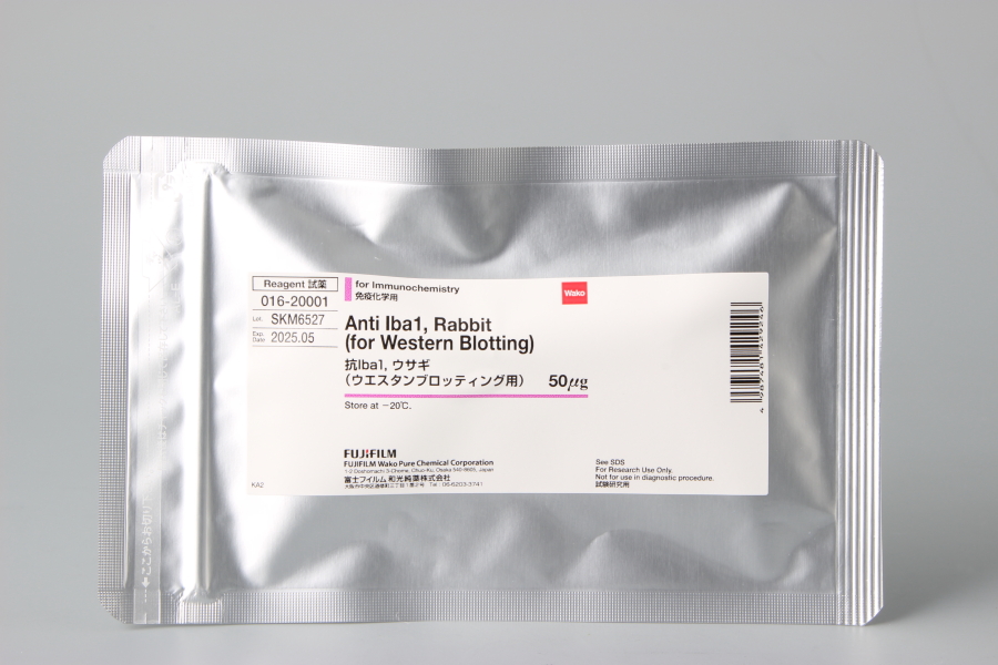
wako Anti Iba1, Rabbit  016-20001 (for Western Blotting)

Iba1: ionized calcium binding adapter molecule 1 小胶质细胞/巨噬细胞特异性蛋白抗体

        钙离子是已知的所有细胞,包括中枢神经系统(CNS)细胞中最重要的信号调解人之一。钙离子通过与各种钙结合蛋白结合发挥信号活动,其中许多钙结合蛋白被划分成一个大的蛋白家族,EF手性蛋白家族。Iba1是一个17 kDa的EF手性蛋白,在巨噬细胞和小胶质细胞中表达,并在这些细胞的活化过程中表达升高。
本抗体以合成的的Iba1羧基末端序列为抗原,该序列是人,大鼠和小鼠Iba1蛋白质保守序列。本抗体(免疫组化用)特异性与小胶质细胞/巨噬细胞反应,免疫双重染色与单克隆抗体结合,可与GFAP单克隆抗体((胶质纤维酸性蛋白,Glial Fibrillary Acidic Protein)联合使用对脑组织和细胞培养物进行双重染色。

特点

S特异性检测小胶质细胞与/巨噬细胞,不与神经细胞与星形胶质细胞发生交叉反应。可与人,大鼠,小鼠的Iba1反应

Wako 016-26721 Anti Iba1, Monoclonal Antibody(NCNP24) for Immunochemistry 50ul
Wako 012-26723 Anti Iba1, Monoclonal Antibody(NCNP24) for Immunochemistry 10ul
Wako 013-26471 Anti Iba1, Rabbit, Red Fluorochrome(635)-conjugated for Immunochemistry 100ul
小胶质细胞/巨噬细胞特异性蛋白抗体(结合红色荧光素635)
Wako 016-26461 Anti Iba1, Rabbit, Biotin-conjugated for Immunochemistry 100ul
小胶质细胞/巨噬细胞特异性蛋白抗体(结合生物素)
Wako  019-19741 Anti Iba1, Rabbit (for Immunocytochemistry) for Immunochemistry 50μg
小胶质细胞/巨噬细胞特异性蛋白抗体(免疫组化)
Wako 016-20001 Anti Iba1, Rabbit (for Western Blotting) for Immunochemistry 50μg
小胶质细胞/巨噬细胞特异性蛋白抗体(免疫印迹)

应用

石蜡切片应用方法:
1. 固定
2. 用0.01M PBS缓冲液清洗二次
3. 使用含有0.3% 过氧化氢的甲醇孵育30分钟
4. 用0.01M PBS缓冲液清洗三次
5. 使用封闭液(含有1.5%的山羊血清和1%BSA的0.001M PBS缓冲液)室温孵育2小时
6. 使用含有Anti Iba1 polyclonal antibody的封闭液4℃孵育过夜
7. 用0.01M PBS缓冲液清洗三次
8. 使用含有生物素标记的抗兔IgG(1:200)的封闭液室温孵育1小时
9. 用0.01M PBS缓冲液清洗三次
10. 使用Elite ABC试剂(含有试剂A1:50,试剂B1:50的0.01M PBS溶液)
11. 用0.01M PBS缓冲液清洗三次

12. 过氧化物酶溶液(含有0.01% 过氧化氢和0.05% DAB的0.05M Tris溶液)

巨噬细胞/小胶质细胞特异性蛋白抗体Iba1的应用

免疫细胞化学⇒和光 Cat. No. 019-19741 (50μg (100μL))
说明书下载地址

http://www.siyaku.com/uh/Flh.do?file_path=/ANTI_FILES/W01/W01W0101-1974/D0010.pdf&now=1481080836480

免疫印迹⇒和光Cat. No. 016-20001 (50μg (100μL))
众所周知,钙离子是中枢神经系统细胞信号传递的重要介质。钙离子通过与各种钙结合蛋白关联激活信号,其中大多数蛋白属于EF手性蛋白家族。
 Iba1属于17-kDa EF蛋白,在巨噬细胞和小胶质细胞中特异性表达,起到正向调节的作用。
和光提供的兔多抗可识别Iba1羧基末端序列的合成肽,这段Iba1蛋白序列在人、大鼠和小鼠中均存在。这些抗体可特异性的识别小胶质细胞和巨噬细胞,适用于脑组织的双重免疫染色,以及细胞培养中与GFAP单抗联合使用(GFAP单抗可特异性的识别星形胶质细胞)。
 
特征:特异性识别人、大鼠、小鼠的小胶质细胞及巨噬细胞,无法作用于神经元和星形胶质细胞。
 
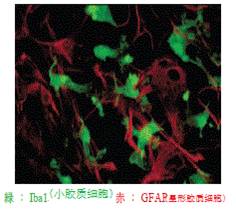
大鼠原代的细胞培养的双重免疫染色和同一位置的对比图像
绿色:Iba1,小胶质细胞/巨噬细胞特异性蛋白抗体作用 (Wako Cat. #019-19741)
红色:星形胶质细胞,与GFAP单抗作用
      (此数据由 日本神经系统科学国家研究所神经化学部提供)

Iba1抗体用于免疫细胞化学(Wako Cat. #019-19741)试验过程

   石蜡切片应用
1)去石蜡
100%二甲苯 10分钟  2次
100%乙醇   5分钟     2次
80%乙醇   5分钟 
70%乙醇   5分钟 
一蒸馏水    5分钟      2次
2)用0.01M缓冲液冲洗两次;
3)在含0.3%过氧化氢的甲醇里孵育30分钟;
4))用0.01M缓冲液冲洗三次;
5)在室温下,放进含有1.5%山羊血清和1%牛血清蛋白的0.001MPBS的封闭液里孵育2小时;
6)在4度下,与 Anti Iba1 Antibody (0.5 μg/mL)(Wako Catalog No. 019‐19741)在封闭液中孵育过夜;
7)用0.01M PBS冲洗3次;
8)在室温下,与生物素标记的anti‐Rabbit IgG Antibody (1:200)在封闭液中孵育1小时;
9)用0.01M PBS冲洗3次;
10)在室温下,与ABC试剂(试剂A(1:50)和试剂B(1:50)与0.01MPBS混匀)孵育1小时
11)用0.01MPBS冲洗3次;
12)在过氧化物酶溶液(含0.01%过氧化氢,0.05%DAB的0.05MTris缓冲液)中孵育;

Anti Iba1, Rabbit (for Immunocytochemistry)   019-19741 介绍

小胶质细胞/巨噬细胞特异性蛋白抗体(免疫组化)

鉴定小胶质细胞的标记物。众多文献引用。产品质量稳定。小胶质细胞属于神经免疫细胞,在当前神经生物学研究的热点。包括从事眼科研究,神经退行性疾病的客户会用到该产品。
文献报道可用于石蜡切片和冰冻切片的免疫组化(IHC)实验。

品牌:Wako
CAS No.:
储存条件:-20℃
纯度:0.5mg/ml

详细介绍:
This product is for research use only. Do not administer it to human.
Antibodies against Macrophage/Microglia-specific Protein Iba1
Calcium ions are known to be one of the most important signal mediators in all cells including central nervous system (CNS) cells. Calcium ions exert their signaling activity through association with various calcium binding proteins, many of which are classified into a large protein family, the EF hand protein family. Iba1 is a 17-kDa EF hand protein that is specifically expressed in macrophages/ microglia and is upregulated during the activation of these cells.Wako has launched rabbit polyclonal antibodies were raised against a synthetic peptide corresponding to the Iba1 carboxy-terminal sequence, which was conserved among human, rat and mouse Iba1 protein sequences. These antibodies are specifically reactive to microglia/ macrophages, are appropriate for immuno-double staining of brain tissues and cell culture in combination with monoclonal antibody to GFAP, which specifically reacts to astrocyte.
Rabbit Anti Iba1 antibody is raised a synthetic peptide corresponding to C-terminus of Iba1.
Preparation: Purified by the antigen affinity chromatography from rabbit antisera and prepared in TBS solution. Contains no preservatives and stabilizers.
Specificity: Specific to microglia and macrophage, but not cross-reactive with neuron and astrocyte. Reactive with human, mouse and rat Iba1.
Working Concentration: Immunocytochemistry 1-2 μg/mL
Storage:Store at -20°C. After opening, aliquot contents and freeze at -20°C.
Package Size: 50 μg (100 μL)
Specificity:
Specific to microglia and macrophage, but not cross-reactive with neuron and astrocyte. Reactive with human, mouse and rat Iba1.
References:参考文献

  1. Imai, Y., Ibata, I., Ito, D., Ohsawa, K. and Kohsaka, S.: Biochem. Biophys. Res. Commun., 224, 855 (1996).
  2. Ito, D., Imai, Y., Ohsawa, K, Nakajima, K, Fukuuchi, Y. and Kohsaka, S.: Brain Res. Mol. Brain Res., 57, 1 (1998).
  3. Ohsawa, K., Imai, Y., Kanazawa, H., Sasaki, Y. and Kohsaka, S.: J. Cell Sci., 113, 3073 (2000).
  4. Sasaki, Y., Ohsawa, K., Kanazawa, H., Kohsaka, S. and Imai, Y.: Biochem. Biophys. Res. Commun., 286, 292 (2001).
  5. Kanazawa, H., Ohsawa, K., Sasaki, Y., Kohsaka, S. and Imai, Y.: J. Biol. Chem., 277, 20026(2002).


iba1小胶质细胞抗体 免疫印迹⇒ 016-20001 (50μg (100μL))

wako Anti Iba1, Rabbit  016-20001 (for Western Blotting)

钙离子作为信号转导通道中的第一信使,在所有细胞包括中枢神经细胞的生命活动中起重要作用。钙离子与不同的钙结合蛋白结合后进行级联反应。Iba1 是能与巨噬细胞和小胶质细胞发生特异性结合的钙结合蛋白,当这些细胞激活后,Iba1 的表达量上调,因此Iba1 通常作为鉴定小胶质细胞的标记物使用。近来研究发现,小胶质细胞除了提供营养、保护神经的作用外,还被证明可产生NO、TNF- α、IL-1 β等物质,因此将小胶质细胞定义为中枢神经系统中的巨噬细胞样吞噬细胞,具有重要的免疫细胞作用。当病原菌,病毒入侵中枢神经系统时会起免疫监视的作用。近来研究发现,该细胞可能与神经退行性疾病的发生和发展有关。本产品是可与小胶质细胞发生特异性反应的兔多抗,配合对星形胶质细胞有特异性的GFAP 单抗,可进行双染色。
抗原:合成肽(Iba1 的C 末端序列)
外观:TBS 溶液(1mg/ml)
提纯:抗原亲和提纯
特异性:对小胶质细胞、巨噬细胞特异反应。不会与神经元、星形胶质细胞发生交叉反应。
能与人、小鼠、大鼠Iba1 反应。
用法: ① Iba1 抗体,兔源(Immunocytochemistry 用)
适合Immunocytochemistry。
只需1 ~ 2 ug/ml 即可进行实验。
② Iba1 抗体,兔源(Western blotting 用)适合Western blotting。
只需0.5 ~ 1ug/ml 即可进行实验。

绿色:Iba1 抗体,兔源(Immunocytochemistry 用)(小胶质细胞)
红色:抗GFAP 抗体(星形胶质细胞)
数据提供:日本国立精神• 神经中心神经研究所代谢研究部

Anti Pax6, Rabbit 抗Pax6抗体,兔源 品牌:Wako


品牌:Wako
CAS No.:
储存条件:-20℃
纯度:
产品编号

(生产商编号)

等级 规格 运输包装 零售价(RMB) 库存情况 参考值

019-27291

for Immunochemistry 10 ul

Anti Pax6, Rabbit                                                      抗Pax6抗体,兔源            品牌:Wako

替代品:016-27343


* 干冰运输、大包装及大批量的产品需酌情添加运输费用


* 零售价、促销产品折扣、运输费用、库存情况、产品及包装规格可能因各种原因有所变动,恕不另行通知,确切详情请联系上海金畔生物科技有限公司。

产品描述相关资料下载相关产品浏览记录 请联系客服

Anti Mouse 4.1G/EPB41L2, Rabbit 抗小鼠4.1G/EPB41L2,兔源 品牌:Wako


品牌:Wako
CAS No.:
储存条件:-20℃
纯度:
产品编号

(生产商编号)

等级 规格 运输包装 零售价(RMB) 库存情况 参考值

018-26421

免疫化学用 50 ul

Anti Mouse 4.1G/EPB41L2, Rabbit                                                       抗小鼠4.1G/EPB41L2,兔源            品牌:Wako


* 干冰运输、大包装及大批量的产品需酌情添加运输费用


* 零售价、促销产品折扣、运输费用、库存情况、产品及包装规格可能因各种原因有所变动,恕不另行通知,确切详情请联系上海金畔生物科技有限公司。

产品描述相关资料下载相关产品浏览记录 请联系客服

Rabbit Apo B-48 ELISA Kit 兔血清载脂蛋白B-48(ApoB-48)ELISA试剂盒 品牌:FUJIFILM Wako Shibayagi


品牌:FUJIFILM Wako Shibayagi
CAS No.:
储存条件:2-10℃
纯度:
产品编号

(生产商编号)

等级 规格 运输包装 零售价(RMB) 库存情况 参考值

628-04901

96 tests 咨询


* 干冰运输、大包装及大批量的产品需酌情添加运输费用


* 零售价、促销产品折扣、运输费用、库存情况、产品及包装规格可能因各种原因有所变动,恕不另行通知,确切详情请联系上海金畔生物科技有限公司。

产品描述相关资料下载相关产品浏览记录 请联系客服

Anti Pax6, Rabbit 抗Pax6抗体,兔源 品牌:Wako


品牌:Wako
CAS No.:
储存条件:-20℃
纯度:
产品编号

(生产商编号)

等级 规格 运输包装 零售价(RMB) 库存情况 参考值

015-27293

for Immunochemistry 50 ul 3,370.00


* 干冰运输、大包装及大批量的产品需酌情添加运输费用


* 零售价、促销产品折扣、运输费用、库存情况、产品及包装规格可能因各种原因有所变动,恕不另行通知,确切详情请联系上海金畔生物科技有限公司。

产品描述相关资料下载相关产品浏览记录 请联系客服

Lactate Dehydrogenase, from Rabbit Muscle (LDH (R.M.)) 品牌:O Yeast


品牌:O Yeast
CAS No.:9001-60-9
储存条件:2-10℃
纯度:
产品编号

(生产商编号)

等级 规格 运输包装 零售价(RMB) 库存情况 参考值

308-50583

50000 units

Lactate Dehydrogenase, from Rabbit Muscle (LDH (R.M.))                                                                品牌:O Yeast


* 干冰运输、大包装及大批量的产品需酌情添加运输费用


* 零售价、促销产品折扣、运输费用、库存情况、产品及包装规格可能因各种原因有所变动,恕不另行通知,确切详情请联系上海金畔生物科技有限公司。

产品描述相关资料下载相关产品浏览记录 请联系客服

Lactate Dehydrogenase, from Rabbit Muscle (LDH (R.M.)) 品牌:O Yeast


品牌:O Yeast
CAS No.:9001-60-9
储存条件:2-10℃
纯度:
产品编号

(生产商编号)

等级 规格 运输包装 零售价(RMB) 库存情况 参考值

306-50584

100000 U

Lactate Dehydrogenase, from Rabbit Muscle (LDH (R.M.))                                                                品牌:O Yeast


* 干冰运输、大包装及大批量的产品需酌情添加运输费用


* 零售价、促销产品折扣、运输费用、库存情况、产品及包装规格可能因各种原因有所变动,恕不另行通知,确切详情请联系上海金畔生物科技有限公司。

产品描述相关资料下载相关产品浏览记录 请联系客服

抗Iba1,兔(用于免疫印迹) Anti Iba1, Rabbit (for Western Blotting)

  • 产品特性
  • 相关资料
  • Q&A
  • 参考文献

抗Iba1,兔(用于免疫印迹)                              Anti Iba1, Rabbit (for Western Blotting)抗Iba1,兔(用于免疫印迹)

Anti Iba1, Rabbit (for Western Blotting)

小胶质细胞标记抗体(WB用)

产品编号

产品名称【中文名称】

规格

包装

016-20001

Anti Iba1, Rabbit (for Western Blotting)

【抗Iba1,兔(用于免疫印迹)】

免疫化学用

50 μg

抗体信息

抗原名

Iba1

适用实验

WB

同种型

IgG

WB图像

抗Iba1,兔(用于免疫印迹)                              Anti Iba1, Rabbit (for Western Blotting)

数据提供:

国立精神神经医疗研究中心 代谢研究部

抗原信息

Iba1C末端序列肽

物种交叉反应性

人,小鼠,大鼠

标签

非标签

抗原别名

AIF-1, IRT1, Protein G1

免疫动物

克隆号

– (多克隆抗体)

详细信息

Iba1是在巨噬细胞/小胶质细胞特异性表达,分子量17000的钙结合蛋白。近年来,小胶质细胞除了对神经营养·保护作用以外,产生的NO、TNF- α、IL-1 β对神经伤害作用很受关注。

本产品用于WB的Iba1抗体特异性检测小胶质细胞。

使用文献

1. Ito, D., Imai, Y., Ohsawa, K., Nakajima, K., Fukuuchi, Y. and Kohsaka, S. : Brain Res.Mol. Brain Res., 57, 1(1998).
  2. Kanazawa, H., Ohsawa, K., Sasaki, Y., Kohsaka, S. and Imai, Y. : J. Biol. Chem., 277, 20026(2002).

实物图

抗Iba1,兔(用于免疫印迹)                              Anti Iba1, Rabbit (for Western Blotting)


欲了解相关资料请点击文字:

Wako神经生物学抗体清单

巨噬细胞/小胶质细胞Iba1抗体


产品编号 产品名称 产品规格 产品等级
016-20001 Anti Iba1, Rabbit (for Western Blotting) 
抗Iba1,兔(用于免疫印迹)
50 μg 免疫化学用

小胶质细胞/巨噬细胞特异性蛋白抗体——Iba1抗体,兔(免疫组化) Anti Iba1, Rabbit (for Immunocytochemistry)

  • 产品特性
  • 相关资料
  • Q&A
  • 参考文献

小胶质细胞/巨噬细胞特异性蛋白抗体——Iba1抗体,兔(免疫组化)小胶质细胞/巨噬细胞特异性蛋白抗体——Iba1抗体,兔(免疫组化)                              Anti Iba1, Rabbit (for Immunocytochemistry)

Anti Iba1, Rabbit (for Immunocytochemistry)


小胶质细胞标记抗体(免疫染色用)

产品编号

产品名称【中文名称】

规格

包装

019-19741

Anti Iba1, Rabbit (for Immunocytochemistry)

【抗Iba1,兔(免疫细胞化学)】

免疫化学用

50 μg

抗体信息

抗原名

Iba1

适用

实验

免疫

染色

同种型

IgG

免疫染色图像

小胶质细胞/巨噬细胞特异性蛋白抗体——Iba1抗体,兔(免疫组化)                              Anti Iba1, Rabbit (for Immunocytochemistry)

抗原信息

Iba1C末端序列肽

物种交叉反应性

人,小鼠,大鼠

标签

非标签

抗原别名

AIF-1, IRT1, Protein G1

免疫

动物

克隆号


(多克隆抗体)

详细信息

Iba1是与巨噬细胞/小胶质细胞特异性表达,分子量17000的钙结合蛋白。近年来,小胶质细胞除了对神经营养·保护作用以外,产生的NO、TNF- α、IL-1 β对神经伤害作用很受关注。

本产品是与小胶质细胞特异性反应的兔多克隆抗体,适用于与星形胶质细胞特异性GFAP单克隆抗体的双重染色。

使用文献

 [1] Jun,et al.(2017).Nature551(7679),232-236.

 [2] Gibson,et al.(2014).Science344(6183),1252304.

 [3] Yan,et al. (2018).Cell, S009286741830285X.

 [4] Pil,et al. (2018). Nature Medicine.

欲了解相关资料请点击文字:

Wako神经生物学抗体清单

巨噬细胞/小胶质细胞Iba1抗体

◆相关资料


Iba1抗体选择指南


小胶质细胞/巨噬细胞特异性蛋白抗体——Iba1抗体,兔(免疫组化)                              Anti Iba1, Rabbit (for Immunocytochemistry)

Iba1抗体系列

参考文献


◆Nature


 1.

Lam, C.K., et al.: Nature, 465, 478(2010).

Embolus extravasation is an alternative mechanism for cerebral microvascular recanalization.

 2.

Stefater, J. A. 3rd. et al.: Nature, 474, 511(2011).   

Regulation of angiogenesis by a non-canonical Wnt-Flt1 pathway in myeloid cells.

 3.

Deng, H. X., et al.: Nature, 477, 211(2011).

Mutations in UBQLN2 cause dominant X-linked juvenile and adult onset ALS and ALS/dementia.

 4.

Lee, Y., et al.: Nature, 487, 433(2012).

Oligodendroglia metabolically support axons and contribute to neurodegeneration.

 5.

Heneka, M. T., et al.: Nature, 493, 674(2013).

NLRP3 is activated in Alzheimer's disease and contributes to pathology in APP/PS1 mice.

 6.

Shao, W., et al.: Nature, 494, 90(2013).

Suppression of neuroinflammation by astrocytic dopamine D2 receptors via αB-crystallin

 7.

Zhang, G., et al.: Nature, 497, 211(2013).

Hypothalamic programming of systemic ageing involving IKK-β, NF-κB and GnRH

 8.

Chung, W. S., et al.: Nature, 504, 394(2013).

Astrocytes mediate synapse elimination through MEGF10 and MERTK pathways.

 9.

Roth, T. L., et al.: Nature, 505, 223(2014).

Transcranial amelioration of inflammation and cell death after brain injury

10.

Najm, F. J., et al.: Nature, 522, 216(2015).

Drug-based modulation of endogenous stem cells promotes functional remyelination in vivo.


11.

Fourgeaud, L., et al.: Nature, 532, 240(2016).

TAM receptors regulate multiple features of microglial physiology.

12.

Vasek, M. J., et al.: Nature, 534, 538(2016).

A complement-microglial axis drives synapse loss during virus-induced memory impairment.


13.

Iaccarino, H. F., et al.: Nature, 540, 230(2016).

Gamma frequency entrainment attenuates amyloid load and modifies microglia.

14.

Bialas, A. R. et al.: Nature, 546, 539(2017).

Microglia-dependent synapse loss in type I interferon-mediated lupus

15.

Mass, E., et al.: Nature, 549, 389(2017).

A somatic mutation in erythro-myeloid progenitors causes neurodegenerative disease.

16.

Jun, J. J., et al.: Nature, 551, 232(2017).

Fully integrated silicon probes for high-density recording of neural activity.

17.

Bussian, T. J., et al.: Nature, 562, 578(2018).
Clearance of senescent glial cells prevents tau-dependent pathology and cognitive decline.

◆CELL


 1.

Lujambio, A., et al.: Cell, 153, 2, 449(2013).

Non-Cell-Autonomous Tumor Suppression by p53.

 2.

Parkhurst, C. N., et al.: Cell, 155, 7, 1596(2013).

Microglia promote learning-dependent synapse formation through brain-derived neurotrophic

factor.

 3.

Wang, Y., et al.: Cell, 160, 6, 1061(2015).

TREM2 Lipid Sensing Sustains the Microglial Response in an Alzheimer’s Disease Model.

 4.

Keren-Shaul, H., et al.: Cell, 169, 7, 1276(2017).

A Unique Microglia Type Associated with Restricting Development of Alzheimer's Disease.

 5.

Ulland, T. K., et al.: Cell, 170, 4, 649(2017).

TREM2 Maintains Microglial Metabolic Fitness in Alzheimer’s Disease.

 6.

Qin, Y., et al.: Cell, 174, 1, 156(2018).

A Milieu Molecule for TGF-β Required for Microglia Function in the Nervous System.

 7.

Yan, S., et al.: Cell, 173, 4, 989(2018).

A Huntingtin Knockin Pig Model Recapitulates Features of Selective Neurodegeneration in

Huntington's Disease

◆Nature Medicine


 1.

Heppner, F. L., et al.: Nat. Med., 2, 146(2005).  

Experimental autoimmune encephalomyelitis repressed by microglial paralysis.

 2.

Nikić, I., et al.: Nat. Med., 4, 495(2011).

A reversible form of axon damage in experimental autoimmune encephalomyelitis and multiple

sclerosis.


 3.

Vom, B. J., et al.: Nat. Med., 12, 1812(2012).

Inhibition of IL-12/IL-23 signaling reduces Alzheimer’s disease–like pathology and cognitive

decline

 4.

Minami, S. S., et al.: Nat. Med., 10, 1157(2014).

Progranulin protects against amyloid β deposition and toxicity in Alzheimer's disease mouse

models.

 5.

Yun, S. P., et al.: Nat. Med., 7, 931(2018).

Block of A1 astrocyte conversion by microglia is neuroprotective in models of Parkinson's disease.

 6.

Mount, C. W., et al.: Nat Med. 5, 572(2018).

Potent antitumor efficacy of anti-GD2 CAR T cells in H3-K27M+ diffuse midline gliomas.


◆Nature Neuroscience


 1.

Zhang, K., et al.: Nat. Neurosci., 10, 1064(2003).

HIV-induced metalloproteinase processing of the chemokine stromal cell derived factor-1 causes

neurodegeneration.

 2.

Ajami, B., et al.: Nat. Neurosci., 12, 1538(2007).

Local self-renewal can sustain CNS microglia maintenance and function throughout adult life

 3.

Mildner, A., et al.: Nat. Neurosci., 12, 1544(2007).

Microglia in the adult brain arise from Ly-6ChiCCR2+ monocytes only under defined host

conditions.

 4.

Bero, A. W., et al.: Nat. Neurosci., 6, 750(2011).

Neuronal activity regulates the regional vulnerability to amyloid-β deposition.

 5.

Fancy, S. P., et al.: Nat. Neurosci., 14, 1009(2011).

Axin2 as regulatory and therapeutic target in newborn brain injury and remyelination.

 6.

Ajami, B., et al.: Nat. Neurosci., 14, 1142(2011).

Infiltrating monocytes trigger EAE progression, but do not contribute to the resident microglia pool.

 7.

Mosher, K. I. et al.: Nat. Neurosci., 11, 1485(2012).

Neural progenitor cells regulate microglia functions and activity.

 8.

Lehmann, S. M., et al.: Nat. Neurosci., 6, 827(2012).

An unconventional role for miRNA: let-7 activates Toll-like receptor 7 and causes

neurodegeneration.


 9.

Kierdorf, K., et al.: Nat. Neurosci., 3, 273(2013).

Microglia emerge from erythromyeloid precursors via Pu.1- and Irf8-dependent pathways

10.

Bialas, A. R. et al.: Nat. Neurosci., 12, 1773(2013).

TGF-β signaling regulates neuronal C1q expression and developmental synaptic refinement

11.

Butovsky, O., et al.: Nat. Neurosci., 1, 131(2014).

Identification of a Unique TGF-β Dependent Molecular and Functional Signature in Microglia.

12.

Saito, T., et al.: Nat. Neurosci., 5, 661(2014).

Single App knock-in mouse models of Alzheimer's disease.

13.

Erny, D., et al.: Nat. Neurosci., 7, 965(2015).

Host microbiota constantly control maturation and function of microglia in the CNS.


14.

Sorge, R. E. et al.: Nat. Neurosci., 8, 1081(2015).

Different immune cells mediate mechanical pain hypersensitivity in male and female mice.

15.

Hama, H., et al.: Nat. Neurosci., 10, 1518(2015).

ScaleS: an optical clearing palette for biological imaging.

16.

Asai, H., et al.: Nat. Neurosci., 11, 1584(2015).

Depletion of microglia and inhibition of exosome synthesis halt tau propagation.

17.

Guan, Z., et al.: Nat. Neurosci., 1, 94(2016).

Injured sensory neuron-derived CSF1 induces microglial proliferation and DAP12-dependent pain.

18.

Grabert, K., et al.: Nat. Neurosci., 3, 504(2016).

Microglial brain region-dependent diversity and selective regional sensitivities to aging

19.

Gonçalves, J. T., et al.: Nat. Neurosci., 6, 788(2016).

In vivo imaging of dendritic pruning in dentate granule cells

20.

Liu, Q., et al.: Nat. Neurosci., 2, 243(2016).

Neural stem cells sustain natural killer cells that dictate recovery from brain inflammation.

21.

Safaiyan, S., et al.: Nat. Neurosci., 8, 995(2016).

Age-related myelin degradation burdens the clearance function of microglia during aging.

22.

Pandya, H., et al.: Nat. Neurosci., 5, 753(2017).

Differentiation of human and murine induced pluripotent stem cells to microglia-like cells

23.

Füger, P., et al.: Nat. Neurosci., 10, 1371(2017).

Microglia turnover with aging and in an Alzheimer's model via long-term in vivo single-cell imaging


◆Nature Immunology


 1.

Wang, Y., et al.: Nat. Immunol., 13, 753(2012).

IL-34 is a tissue-restricted ligand of CSF1R required for the development of Langerhans cells and

microglia.

 2.

Goldmann, T., et al.: Nat. Immunol., 17, 797(2016).

Origin, fate and dynamics of macrophages at central nervous system interfaces


 3.

Haimon, Z., et al.: Nat. Immunol., 19, 636(2018).

Re-evaluating microglia expression profiles using RiboTag and cell isolation strategies.

◆Nature Biotechnology


 1.

Park, S. I., et al.: Nat. Biotechnol., 33, 1280(2015).

Soft, stretchable, fully implantable miniaturized optoelectronic systems for wireless optogenetics

 2.

Staahl, B. T., et al.: Nat. Biotechnol., 35, 431(2017).

Efficient genome editing in the mouse brain by local delivery of engineered Cas9 ribonucleoprotein

complexes

◆Nature Methods


 1.

Clark, J. J., et al.: Nat. Methods., 7, 126(2010).

Chronic microsensors for longitudinal, subsecond dopamine detection in behaving animals

 2.

Prevedel, R., et al.: Nat. Methods., 13, 1021(2016).
Fast volumetric calcium imaging across multiple cortical layers using sculpted light

◆Neuron


 1.

Simard, A. R., et al.: Neuron, 49, 4, 489(2006).

Bone marrow-derived microglia play a critical role in restricting senile plaque formation in

Alzheimer's disease.

 2.

Bhaskar, K., et al.: Neuron, 68, 1, 19(2010).

Regulation of tau pathology by the microglial fractalkine receptor.

 3.

Bergmann, O., et al.: Neuron, 74, 4, 634(2012).

The Age of Olfactory Bulb Neurons in Humans

 4.

Schafer, D. P., et al.: Neuron, 74, 4, 691(2012).

Microglia sculpt postnatal neural circuits in an activity and complement-dependent manner.

 5.

Paolicelli, R. C., et al.: Neuron, 95, 2, 297(2017).

TDP-43 Depletion in Microglia Promotes Amyloid Clearance but Also Induces Synapse Loss.

 6.

Tufail, Y., et al.: Neuron, 93, 3, 574(2017).

Phosphatidylserine Exposure Controls Viral Innate Immune Responses by Microglia.

 7.

Abud, E. M., et al.: Neuron, 94, 2, 278(2017).

iPSC-Derived Human Microglia-like Cells to Study Neurological Diseases.

 8.

Bohlen, C. J., et al.: Neuron, 94, 4, 759(2017).

Diverse Requirements for Microglial Survival, Specification, and Function Revealed by

Defined-Medium Cultures.

 9.

De, Biase, L. M., et al.: Neuron, 95, 2, 341(2017).

Local Cues Establish and Maintain Region-Specific Phenotypes of Basal Ganglia Microglia.

10.

Hwang, H. W., et al.: Neuron, 95, 6, 1334(2017).

cTag-PAPERCLIP Reveals Alternative Polyadenylation Promotes Cell-Type Specific Protein Diversity

and Shifts Araf Isoforms with Microglia Activation.

11.

Lehrman, E. K., et al.: Neuron, 100, 1, 120(2018).

CD47 Protects Synapses from Excess Microglia-Mediated Pruning during Development.

12.

López-Erauskin, J., et al.: Neuron, 100, 4, 816(2018).

ALS/FTD-Linked Mutation in FUS Suppresses Intra-axonal Protein Synthesis and Drives Disease

Without Nuclear Loss-of-Function of FUS

产品编号 产品名称 产品规格 产品等级
019-19741 Anti Iba1, Rabbit (for Immunocytochemistry) 
抗Iba1,兔(免疫细胞化学)
50 μg 免疫化学用

抗人类NAIP,兔 Anti Human NAIP, Rabbit

  • 产品特性
  • 相关资料
  • Q&A
  • 参考文献

抗人类NAIP,兔                              Anti Human NAIP, Rabbit抗人类NAIP,兔

Anti Human NAIP, Rabbit

凋亡相关因子NAIP抗体

产品编号

产品名称【中文名称】

规格

包装

019-24251

Anti Human NAIP, Rabbit

【抗人类NAIP,兔】

免疫化学用

20 μL

抗体信息

抗原名

NAIP

适用实验

WB

同种型

IgG

WB图像

(NAIP表达细胞(FLT8)提取物)

抗人类NAIP,兔                              Anti Human NAIP, Rabbit

抗原信息

人NAIP重组

物种交叉反应性

标签

非标签

抗原别名

Neuronal Apoptosis Inhibitory Protein, BIRC1, NLRB1, psiNAIP

免疫动物

克隆号

– (多克隆抗体)

详细信息

Neuronal Apoptosis Inhibitory Protein(NAIP)是抑制凋亡的蛋白质之一。据报道称 NAIP 是Inhibitor of Apoptosis Proteins(IAP)的家族成员,选择性抑制氧化应激细胞凋亡。另外,关于 NAIP 与神经变性疾病(阿尔茨海默病、多发性硬化症)病因相关的报道很多。

本产品是识别人 NAIP 抗体。

使用文献


产品编号 产品名称 产品规格 产品等级
019-24251 Anti Human NAIP, Rabbit 
抗人类NAIP,兔
20 μL 免疫化学用

JNK2 (56G8) 兔单克隆抗体 JNK2 (56G8) Rabbit mAb

JNK2 (56G8) 兔单克隆抗体

JNK2 (56G8) Rabbit mAb

详细描述:
JNK2 (56G8) Rabbit mAb 兔单抗能检测内源性的总JNK2蛋白。这个抗体不能检测JNK1 或 JNK3 蛋白。该单克隆抗体是采用人源 JNK2/MBP融合蛋白免疫动物而获得。JNK2 (56G8) Rabbit mAb detects endogenous levels of total JNK2 protein. This antibody does not recognize JNK1 or JNK3 protein.Monoclonal antibody is produced by immunizing animals with a human JNK2/MBP fusion protein.

应用范围:W; 反应种属:Human,Mouse,Rat,Hamster,Monkey,Mink; 灵敏度:Endogenous; MW (kDa):46, 54; Isotype:Rabbit; 标记:无标记。

货号 产品名称 品牌 购买
货号 名称 单位 购买
9258T JNK2 (56G8) 兔单克隆抗体 20 µl 咨询客服
9258S JNK2 (56G8) 兔单克隆抗体 200µl 咨询客服

BETA-ACTIN 兔单抗 β-Actin Rabbit Monoclonal Antibody

BETA-ACTIN 兔单抗

β-Actin Rabbit Monoclonal Antibody

详细描述:
β-Actin primary antibodies can be used for detection of β-Actin by Western blot or by In-Cell Western™ assay. These primary antibodies can be used as a normalization antibody when performing two-color detection.

应用:WB,IN-Cell-Western;反应种属:Human, mouse, rat, monkey;抗体来源及亚型:Rabbit IgG;检测抗原分子量大小:45 kDa;无标记。

货号 产品名称 品牌 购买
货号 名称 单位 购买
926-42210 BETA-ACTIN 兔单抗 100 µL 咨询客服

TNFRSF13C/BAFF-R (E3C1Q) XP® 兔单克隆抗体 TNFRSF13C/BAFF-R (E3C1Q) XP® Rabbit mAb

TNFRSF13C/BAFF-R (E3C1Q) XP® 兔单克隆抗体

TNFRSF13C/BAFF-R (E3C1Q) XP® Rabbit mAb

详细描述:

https://media.cellsignal.com/pdf/62518.pdf

货号 产品名称 品牌 购买
货号 名称 单位 购买
62518S TNFRSF13C/BAFF-R (E3C1Q) XP® 兔单克隆抗体 100ul 咨询客服
62518T TNFRSF13C/BAFF-R (E3C1Q) XP® 兔单克隆抗体 20ul 咨询客服

BETA-TUBULIN 兔单抗 β-Tubulin Rabbit Polyclonal Antibody 

BETA-TUBULIN 兔单抗

β-Tubulin Rabbit Polyclonal Antibody 

详细描述:
α or β-Tubulin primary antibodies can be used for detection of α or β-Tubulin by Western blot or by In-Cell Western™ assay. These primary antibodies can be used as a normalization antibody when performing two-color detection.

应用:WB,IN-Cell-Western;反应种属:Human, mouse, rat, monkey, bovine;抗体来源及亚型:Rabbit IgG;检测抗原分子量大小:55 kDa;无标记。

货号 产品名称 品牌 购买
货号 名称 单位 购买
926-42211 BETA-TUBULIN 兔单抗 100 µL 咨询客服

TNFRSF13C/BAFF-R (E3C1Q) XP® 兔单克隆抗体 TNFRSF13C/BAFF-R (E3C1Q) XP® Rabbit mAb

TNFRSF13C/BAFF-R (E3C1Q) XP® 兔单克隆抗体

TNFRSF13C/BAFF-R (E3C1Q) XP® Rabbit mAb

详细描述:

https://media.cellsignal.com/pdf/62518.pdf

货号 产品名称 品牌 购买
货号 名称 单位 购买
62518S TNFRSF13C/BAFF-R (E3C1Q) XP® 兔单克隆抗体 100ul 咨询客服
62518T TNFRSF13C/BAFF-R (E3C1Q) XP® 兔单克隆抗体 20ul 咨询客服

MyoD1 (D8G3) XP® 兔单克隆抗体 MyoD1 (D8G3) XP® Rabbit mAb 

MyoD1 (D8G3) XP® 兔单克隆抗体

MyoD1 (D8G3) XP® Rabbit mAb 

详细描述:
MyoD1 (D8G3) XP® Rabbit mAb recognizes endogenous levels of total MyoD1 protein.Monoclonal antibody is produced by immunizing animals with a synthetic peptide corresponding to residues surrounding Gly190 of human MyoD1 protein.

应用范围:W IF-IC; 反应种属:Human; 灵敏度:Endogenous; MW (kDa):45; Isotype:Rabbit IgG; 标记:无标记。

货号 产品名称 品牌 购买
货号 名称 单位 购买
13812T MyoD1 (D8G3) XP® 兔单克隆抗体 20 µl  商城订购
13812S MyoD1 (D8G3) XP® 兔单克隆抗体 100µl 商城订购

PGRMC1 (D6M5M) XP® 兔单克隆抗体  PGRMC1 (D6M5M) XP® Rabbit mAb

PGRMC1 (D6M5M) XP® 兔单克隆抗体 

PGRMC1 (D6M5M) XP® Rabbit mAb

详细描述:
PGRMC1 (D6M5M) XP® Rabbit mAb recognizes endogenous levels of total PGRMC1 protein. This antibody does not cross-react with PGRMC2 protein.Monoclonal antibody is produced by immunizing animals with a synthetic peptide corresponding to residues near the amino terminus of human PGRMC1 protein.

应用范围:W IP IHC-P IF-IC; 反应种属:Human,Mouse,Rat,Monkey; 灵敏度:Endogenous; MW (kDa):25; Isotype:Rabbit IgG; 标记:无标记。

货号 产品名称 品牌 购买
货号 名称 单位 购买
13856T PGRMC1 (D6M5M) XP® 兔单克隆抗体  20 µl  商城订购
13856S PGRMC1 (D6M5M) XP® 兔单克隆抗体  100µl 商城订购

Integrin α1/CD49a (E9K2J) XP® 兔单克隆抗体 Integrin α1/CD49a (E9K2J) XP® Rabbit mAb

Integrin α1/CD49a (E9K2J) XP® 兔单克隆抗体

Integrin α1/CD49a (E9K2J) XP® Rabbit mAb

详细描述:

https://media.cellsignal.com/pdf/15574.pdf

货号 产品名称 品牌 购买
货号 名称 单位 购买
15574S Integrin α1/CD49a (E9K2J) XP® 兔单克隆抗体 100ul 咨询客服

COX IV 兔单抗 COX IV Rabbit Monoclonal Antibody

COX IV 兔单抗

COX IV Rabbit Monoclonal Antibody

详细描述:
The COX IV Rabbit Monoclonal Primary Antibody can be used for detection of COX IV by Western blot, or as a normalization antibody when performing two-color detection. Its target molecular weight is 17 kDa.

应用:WB,IN-Cell-Western;反应种属:Human, rabbit, monkey, zebrafish, bovine, pig;抗体来源及亚型:Rabbit IgG;检测抗原分子量大小:17kDa;无标记。

货号 产品名称 品牌 购买
货号 名称 单位 购买
926-42214 COX IV 兔单抗 100 µL 咨询客服