细胞剥离/解离溶液

  • 产品特性
  • 相关资料
  • Q&A
  • 参考文献

细胞剥离/解离溶液细胞剥离/解离溶液


  请用于剥离粘附细胞或分散各种组织的细胞。

  质量测试:外观、渗透压、pH、支原体测试、无菌测试、实际应用测试、病毒测试*3

  *3:使用已通过猪细小病毒测试的胰蛋白酶(1:250)。

相关产品


  以下产品是从 Clostridium histolyticum(溶组织梭菌) 提取的胶原酶。


产品编号

产 品 名 称

规 格

容 量

038-22361
034-22363
032-22364

Collagenase
胶原酶

用于分散细胞

100 mg
  1 g
  5 g

031-17601
037-17603
035-17604

Collagenase Type !
对肺、上皮组织、脂肪组织的用于分散细胞效果显著。

用于分散细胞

100 mg
500 mg
  1 g

038-17851
032-17854

Collagenase Type V
对胰脏的用于分散细胞效果显著。

用于分散细胞

100 mg
  1 g

035-17861
031-17863
039-17864

Collagenase Type X
对肝脏、心脏、胸腺、唾液腺的用于分散细胞效果显著。

用于分散细胞

100 mg
  500 mg
  1 g


产品编号 产品名称 产品规格 产品等级
201-18841 0.25w/v% Trypsin Solution with Phenol Red 100ml 用于培养细胞
202-16931 0.05w/v% Trypsin-0.53mmol/l EDTA・4Na Solution with Phenol Red 100ml 用于培养细胞
204-16935 0.05w/v% Trypsin-0.53mmol/l EDTA・4Na Solution with Phenol Red 100ml 用于培养细胞
209-16941 0.25w/v% Trypsin-1mmol/l EDTA・4Na Solution with Phenol Red 100ml 用于培养细胞
201-16945 0.25w/v% Trypsin-1mmol/l EDTA・4Na Solution with Phenol Red 500ml 用于培养细胞
208-17251 0.5w/v% Trypsin-5.3mmol/l EDTA・4Na Solution without Phenol Red(×10) 100ml 用于培养细胞
206-17291 0.5w/v% Trypsin-5.3mmol/l EDTA・4Na Solution with Phenol Red(×10) 100ml 用于培养细胞

Wako 神经细胞用分散液

  • 产品特性
  • 相关资料
  • Q&A
  • 参考文献

Wako 神经细胞用分散液神经细胞用分散液

可简单回收神经细胞


  本产品可以从大鼠/小鼠的中枢神经组织中分散、回收神经细胞。本产品由3种溶液(酶溶液、分散液、去除液)组成,无需调制,在保持细胞高生存率状态下可简单地对神经细胞进行回收。

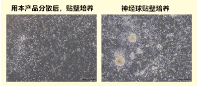
  使用本产品分散神经球后,贴壁培养时死细胞少,可以在易于观察的状态下培养神经细胞。


◆特点


● 可简单、稳定地从中枢神经细胞或神经球中回收神经细胞;

● 即用型


数据


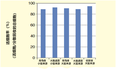
● 冻存的神经细胞分散时的活细胞存活率


Wako 神经细胞用分散液


细胞名称

海马体,小鼠(胎生16日)来源

总细胞数(cells/vial)

4.32×10⁵

活细胞数(cells/vial)

3.38×10⁵

活细胞率(%)

89

大脑皮质,小鼠(胎生15日)来源

总细胞数(cells/vial)

6.64×10⁶

活细胞数(cells/vial)

6.09×10⁶

活细胞率(%)

92

海马体,大鼠(胎生19日)来源

总细胞数(cells/vial)

1.37×10⁶

活细胞数(cells/vial)

1.24×10⁶

活细胞率(%)

90

大脑皮质,大鼠(胎生17日)来源

总细胞数(cells/vial)

1.32×10⁷

活细胞数(cells/vial)

1.17×10⁷

活细胞率(%)

89

纹状体,大鼠(胎生17日)来源

总细胞数(cells/vial)

2.73×10⁶

活细胞数(cells/vial)

2.58×10⁶

活细胞率(%)

95

把各脑组织解冻,使用本品对细胞进行分散、回收,确认细胞活率。

可以确认使用本产品从冻存脑组织中回收细胞的活率可达约90%


◆神经球的分散


Wako 神经细胞用分散液


  正常人 iPS 细胞形成的神经球,贴壁培养,诱导分化为神经细胞。

  使用本产品后进行神经球的贴壁培养,比较发现死细胞更少,可以在容易观察神经细胞的状态情况下进行培养。

  (数据提供:东京慈惠会医科大学 再生医学研究部 冈野 JAMES 洋尚先生、田原 麻由,东京慈惠会医科大学小儿科学讲座 日暮 宪道先生)


◆试剂盒内容

神经细胞用分散液

神经细胞用分散液 S
酶溶液

5.0 mL × 4 支

2.5 mL × 10 支

分散液

5.0 mL × 4 支

2.5 mL × 10 支

去除液

5.0 mL × 4 支

2.5 mL × 10 支


◆产品列表


产品编号

产品名称

产品规格

包装

291-78001

Neuron Dissociation Solutions

神经细胞用分散液

细胞培养用

4 Set

297-78101

Neuron Dissociation Solutions S

神经细胞用分散液S

细胞培养用

10 Set


◆相关产品


神经细胞用培养基


产品编号

产品名称

产品规格

包装

148-09671

Neuron Culture Medium

神经细胞用培养基

细胞培养用

100 mL


产品编号 产品名称 产品规格 产品等级

iStock 人来源细胞冻存液

  • 产品特性
  • 相关资料
  • Q&A
  • 参考文献

人来源细胞冻存液iStock                              人来源细胞冻存液

iStock

iStock                              人来源细胞冻存液


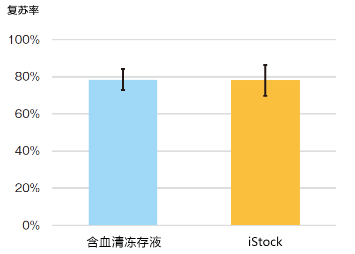
比较LYMPHOTEC目前使用的含血清冻存液与iStock的性能


iStock                              人来源细胞冻存液


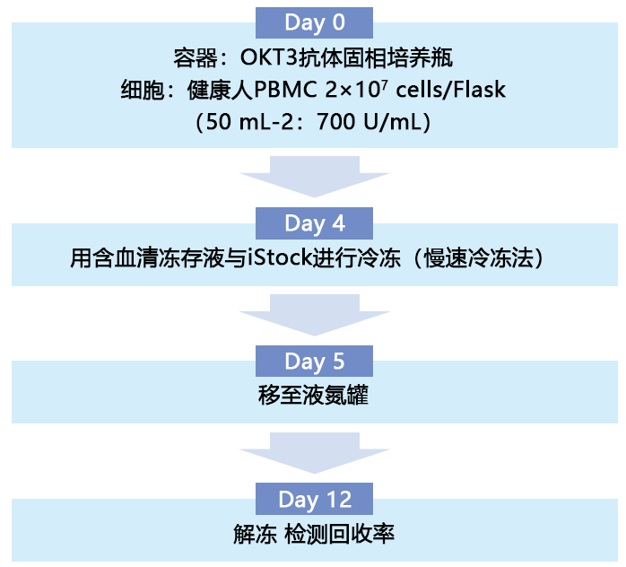
从 9 名知情同意的健康者血液中,分离出外周血单核细胞(PBMC),并使用OKT3抗体固相培养瓶和LYMPHOTEC本公司的淋巴细胞培养基培养4天。使用含血清冻存液和iStock,将增殖的细胞制备成1.5×10⁷ cells/mL,采用慢速冷冻法,冷冻于-80℃。第二天,转移至液氮罐中,保存7天后解冻,比较回收率(解冻后细胞数/冷冻前细胞数×100%)。

iStock使用方法


iStock                              人来源细胞冻存液


◆规格


适用细胞:◯人免疫细胞     ◯人间充质干细胞      ◯人iPS细胞     

保存条件:冷藏、避光、2-10℃

无菌检查:◯内毒素:显色法    ◯支原体:培养法    ◯真菌、细菌:琼脂平板表面涂抹法


操作步骤:

1. 细胞冻存   

❶将细胞移至离心管。

❷离心分离(800×g左右,3-5 min),用抽吸器去除上清。向~1.5×10⁷个细胞中加入1 mL的 iStock,缓慢地进行移液。

❸将细胞悬液分注至冷冻管中。

❹在-80℃的低温冷冻箱进行冻存(根据细胞类型,将冷冻管转移至市售的冷冻容器进行冻存)。

❺第二天移至液氮罐中。


iStock                              人来源细胞冻存液


2. 细胞解冻    

❶在37°C的温水浴中解冻冷冻管。

❷立即与约10 mL的培养液混合。

❸离心分离(800×g左右,3-5 min),用抽吸器去除上清。

❹在适量的培养液中悬浮,转移至培养容器,开始培养。


注意事项:

◯请勿用于人体。

◯本产品未获得医药用品的相关许可证明。

◯在使用前,先对使用的细胞进行确认测试。

◯对于因本产品的使用问题造成的任何事故或损坏,本公司概不负责。

◯使用上如有疑问,请联系(株)LYMPHOTEC。

点击此处下载产品宣传页


产品编号 产品名称 产品规格 产品等级
385-19451 iStock 120 mL

CultureSure无血清细胞冻存液 通用动物细胞的冻存液

  • 产品特性
  • 相关资料
  • Q&A
  • 参考文献

通用动物细胞的冻存液CultureSure无血清细胞冻存液                              通用动物细胞的冻存液

CultureSure无血清细胞冻存液


  本品是适用于冻存动物细胞的无血清细胞通用冻存液。

  利用缓慢冻存法冻存细胞,细胞复苏活率高。

  另外,本品含有BSA和DMSO。


◆特点


● 是具有与含血清产品同等功能的无血清冻存液

● 能通过缓慢冷冻法进行冻存

● 可长期存放在-80℃

● 无需配制试剂


质量检测项目

● 无菌检测

● 内毒素检测

● 支原体检测


使用方法


1.    冻存

  ① 用试管收集细胞。

  ② 离心去除上清。

  ③ 往试管中加入本品,重悬细胞。

  ④ 将悬浮液分注到保存用试管。

  ⑤ 将保存用试管在-80℃中冻存一晚。

  ⑥ 存放于-150℃或-80℃。


2.    细胞复苏

  ① 用37℃的水浴锅解冻冻存的试管。

  ② 在培养使用的培养基中进行悬浮。

  ③ 离心去除上清后在培养基中进行重悬。

  ④ 细胞铺板。


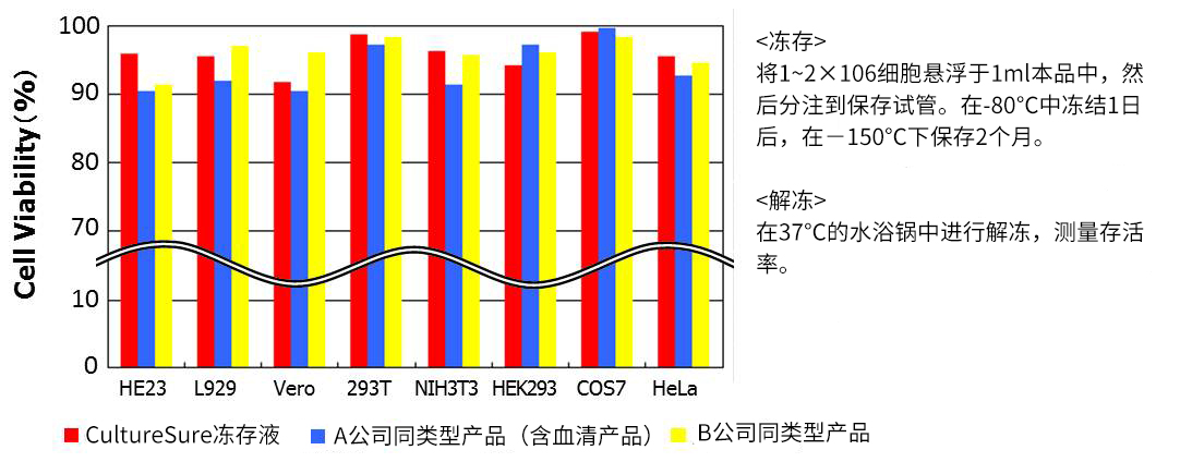
冻存数据①:-150℃,2个月


CultureSure无血清细胞冻存液                              通用动物细胞的冻存液

★维持与含血清冻存液同样的细胞存活率。


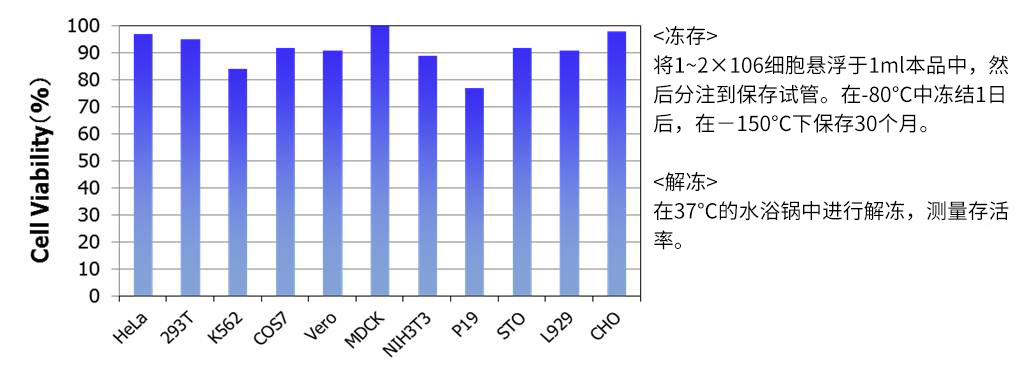
冻存数据②:-150℃,30个月

CultureSure无血清细胞冻存液                              通用动物细胞的冻存液

★细胞保存长达30个月仍保持高存活率


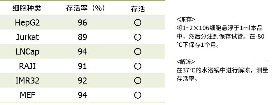
冻存数据③:-80℃,1个月


CultureSure无血清细胞冻存液                              通用动物细胞的冻存液


产品列表

产品编号

产品名称

等级

包装

039-23511

CultureSure Freezing Medium
CultureSure无血清通用细胞冻存液

细胞培养用

100 mL


相关产品


★液体培养基・细胞培养用试剂★

液体培养基、平衡盐溶液、细胞剥离・分散用溶液、抗生素,细胞外基质等产品陆续上市。

★StemSure® 冻存液★

适合冻存小鼠ES细胞或人iPS细胞的细胞保存溶液。

产品编号

产品名称

等级

包装

195-16031

StemSure Freezing Medium
StemSure干细胞冻存液

细胞培养用

100 mL


以上产品仅供实验・研究使用,不可用作“医药品”、“食品”或“家庭用品”。


更多产品资料请点击文字:CultureSure® 无血清细胞冻存液

欢迎下载电子版,或联系客服或当地经销商索取纸质版单页


参考文献

1.

Kusakisako, K., Masatani, T., Yada, Y., Talactac, M. R., Hernandez, E. P., Maeda, H., … & Tanaka, T. (2016). Improvement of the cryopreservation method for the Babesia gibsoni parasite by using commercial freezing media. Parasitology international, 65(5), 532-535. 全文

2.

Shirozu, T., Soga, A., & Fukumoto, S. (2020). Identification and validation of a commercial cryopreservation medium for the practical preservation of Dirofilaria immitis microfilaria. 全文

3.

Lai, Y. C., Ushio, N., Rahman, M. M., Katanoda, Y., Ogihara, K., Naya, Y., … & Sunaga, T. (2018). Aberrant expression of microRNAs and the miR‐1/MET pathway in canine hepatocellular carcinoma. Veterinary and comparative oncology, 16(2), 288-296. 全文

4.

Rahman, M. M., Lai, Y. C., Husna, A. A., Chen, H. W., Tanaka, Y., Kawaguchi, H., … & Miura, N. (2019). Aberrantly expressed snoRNA, snRNA, piRNA and tRFs in canine melanoma. Veterinary and Comparative Oncology.  全文

产品编号 产品名称 产品规格 产品等级

CryoScarless ® DMSO-Free 高存活率细胞冻存液

  • 产品特性
  • 相关资料
  • Q&A
  • 参考文献

CryoScarless ® DMSO-Free                              高存活率细胞冻存液

高存活率细胞冻存液

CryoScarless® DMSO-Free

CryoScarless® DMSO-Free 是一款不含蛋白和 DMSO,适用于多种细胞的细胞冻存液。各种细胞在解冻后具有很高的培养活率,且维持了干细胞的多分化性(未分化状态)。


※ 本产品仅供科研,不可用于科研外其他用途。

关于含有DMSO冻存液的问题

DMSO作为普通细胞冷冻保护成分常被添加到冻存液中,但同时也是对人体有害的成分,且极易被皮肤吸收。除具有细胞毒性外,如同下列论文所述,是影响细胞分化的因子之一。因此,为获得更精确的实验结果,无DMSO的细胞保存是必须的。

● 使用DMSO将人骨髓白血病细胞株 HL-60 分化为粒细胞。1

● 使用DMSO将小鼠间充质干细胞分化为心肌细胞。 2

● 使用含有DMSD的冻存液冻存人ES细胞发现未分化标记Oct-4的表达降低。 3


参考文献


1. Jiang, G., et al., Int. Immunopharmacol.(7), 1204~1213 (2006).

2. Young, D. A., et al., Biochem. Biophys. Res. Commun.322 (3), 759~765 (2004).

3. Katkov, I. I., et al., Cryobiology, 53 (2), 194~205 (2006).

◆特点


● 不含血清和蛋白,因此不受白蛋白和球蛋白等蛋白的影响。

● 不受DMSO毒性和蛋白影响,可安全且高活率地冻存细胞。

● 大部分细胞在冻存后复苏,存活率可达90%以上。

● 已有人/小鼠的正常细胞、肿瘤细胞株、干细胞等各种细胞的冻存应用。

● 冻存后可维持干细胞的分化性能。

● 同样适用于无血清培养。

● 经无菌测试确认不含细菌、真菌与支原体污染。

● 本产品可在4°C下长期保存。(有效期2年)。 


※ 使用台盼蓝处理CryoScarless保存的细胞时,请务必清洗细胞。

◆应用实例1


大鼠间充质干细胞分化能力


CryoScarless ® DMSO-Free                              高存活率细胞冻存液


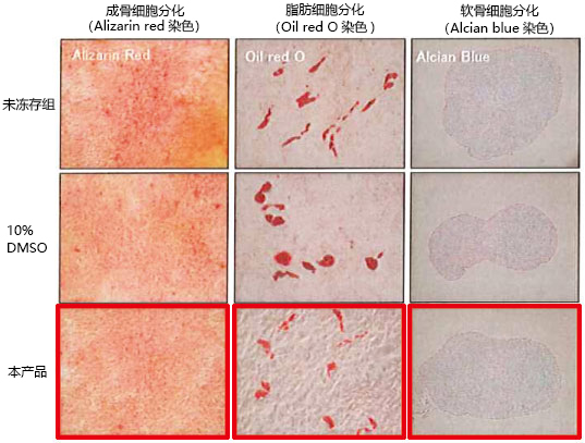
确认使用本产品冻存的大鼠间充质干细胞在复苏后的分化能力,并与未冻存组以及10% DMSO保存的细胞进行比较。结果显示,使用本产品冻存的细胞皆维持良好的分化性能。

大鼠间充干细胞的存活率


CryoScarless ® DMSO-Free                              高存活率细胞冻存液


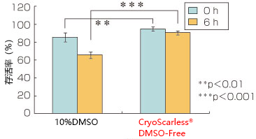
检测使用本产品冻存(1周)的大鼠间充质干细胞在复苏后的存活率以及 6 h后的存活率,并与10% DMSO保存的情况进行比较。本产品无论是在刚复苏(0 h),还是贴壁后(6 h)皆显示更高的存活率。

◆应用实例2


使用 CryoScarless® DMSO-Free 简单冻存后的小鼠 ES 细胞


实验方法:


将小鼠 ES 细胞(FKHR+/+)在明胶包被的96孔板上于37°C培养48 h后,进行简单地冻存。吸走培养基并用PBS清洗,然后分别添加10%DMSO/培养基,或CryoScarless® DMSO-Free,在-80°C下保存。24 h后迅速复苏,添加加热至37°C的培养基,在37°C下继续培养。6 h后使用光学显微镜观察相衬图像。


CryoScarless ® DMSO-Free                              高存活率细胞冻存液

结果:


可观察到在10%DMSO/培养基中保存的小鼠ES细胞在复苏后活细胞减少(左图),而在 CryoScarless® DMSO-Free 中保存的小鼠ES细胞状态得到改善,可观察到大部分的活细胞。

讨论:


在对多个转基因克隆的培养板一起进行冻存时(简易冻存),使用CryoScarless® DMSO-Free 更加方便。利用 CryoScarless® DMSO-Free 进行冻存,存活细胞更多,更易于未分化小鼠ES细胞的传代培养(在一定细胞密度下每2天传代一次),并可顺利进行分化实验。

数据提供


熊本大学发育医学研究所 干细胞部门 组织干细胞领域 田村洁美老师


CryoScarless ® DMSO-Free                              高存活率细胞冻存液

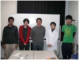
小川峰太郎老师(组织干细胞领域教授:中间),田村洁美老师(右起2)与研究室成员


◆应用实例3


对于人牙髓来源间充质干细胞的体内实验,CryoScarless® DMSO-Free 拥有更出色的冻存能力


日本牙科大学生命牙学部 发育、再生医科讲座 中原贵教授

生命牙科学讲座 望月 真衣 助教


Establishment of xenogeneic serum-free culture methods for handling human dental pulp stem cells using clinically oriented in-vitro and in-vivo conditions.

Stem Cell Research & Therapy,9:25, (2015).


背景


由于再生医学不断普及,因此急需确立可安全地培养从患者获得的间充质干细胞的方法。近年来,通过牙科治疗从拔牙的牙组织(牙髓)中获得的间充质干细胞,显示比骨髓干细胞高3~4倍的高增殖性,且没有伴随染色体异常的细胞变化。而且牙髓干细胞拥有与骨髓干细胞相同的多分化性能。由于在体外培养可大量增殖,灵活利用低癌变和成瘤风险的牙髓干细胞,对于确保医疗安全性的再生医学来说是一个极具吸引力的选择。

作为再生医学中必不可少的"细胞冻存",已经证明CryoScarless® DMSO-Free(BioVerde公司)是一款优秀的细胞冻存液。换言之,使用该细胞冻存液冻存的人牙髓干细胞拥有与未经冻存培养的牙髓干细胞相同的增殖能与多分化能力,且未发现染色体异常。

本文着眼于牙髓干细胞活跃的增殖能力,并证实了CryoScarless® DMSO-Free不会影响细胞增殖能力。

方法及结果


从20~37岁成人拔下来的智齿分离牙髓细胞并进行无血清培养,培养至传代数为1时使用CryoScarless® DMSO-Free冻存。3个月后再次培养冻存的细胞,传代数到3时进行以下的细胞评价。同时,对未经冻存的牙髓细胞也进行了相同的评价。

使用相差显微镜观察细胞形态,确认纺锤形的成纤维细胞形态没有发生变化(图-1)。根据增殖曲线分析的结果,经过12天的培养,呈现一致的细胞增殖模式,两者间在统计学上的没有显著差异(图-2)。另外,通过Bromodeoxyuridine(BrdU)进行细胞分裂能力分析的结果显示,可观察到对数生长期内大量的BrdU阳性细胞,细胞数在统计学上没有显著差异(图-3)。

CryoScarless ® DMSO-Free                              高存活率细胞冻存液

图-1 人牙髓干细胞的相差显微镜图像

无论CryoScarless® DMSO-Free冻存(左)还是未经冻存(右)的细胞都呈现出相同的成纤维细胞样形态。

CryoScarless ® DMSO-Free                              高存活率细胞冻存液

图-2 人牙髓干细胞增殖曲线分析


细胞培养期间,未发现两者在统计学上的显著差异。

CryoScarless ® DMSO-Free                              高存活率细胞冻存液

图-3 人牙髓干细胞在对数生长期内的BrdU分析

可观察到大部分细胞摄入BraU(绿)(左),未发现两者的BraU阳性细胞数在统计学上的显著差异(右)。

结论


除了本文中报告的细胞增殖能力评价之外,使用CryoScarless® DMSO-Free冻存的牙髓干细胞不仅维持了成骨细胞、脂肪细胞、软骨细胞的多分化能力,也能维持向免疫缺陷小鼠的皮下移植并引起硬组织形成的能力。并且该产品并不影响干细胞的特性。

另外,值得注意的是,使用CryoScarless® DMSO-Free冻存的牙髓干细胞即使长期培养至传代数为10后仍然保持染色体的二倍体和正常核型,大量培养的细胞中也没有发现染色体异常。

在再生医学中必不可少的"细胞冻存"中,作为冷冻保护剂使用的Dimethyl sulfoxide(DMSO)可能会有细胞毒性和引起意外的细胞分化,因此,我们十分期待能够确立一个像该产品一样的无DMSO的细胞冻存体系。

本报道中使用易于获得的牙髓来源间充质干细胞,研究了CryoScarless® DMSO-Free对干细胞特性的影响,并确认了使用该产品冻存的细胞与未经冻存的牙髓干细胞拥有相同的细胞特性。因此,CryoScarless® DMSO-Free在间充质干细胞基础研究以及临床前研究,甚至是将来的再生医学领域中,有望成为一款可以提供安全且高预测性数据和治疗成果的细胞冻存液。

CryoScarless ® DMSO-Free                              高存活率细胞冻存液


中原贵教授(前排右),望月真衣助教(前排左)与研究室成员

◆实际应用的细胞和冻存效果


细胞株

存活率(%)*

L929

小鼠成纤维细胞

97.5 ± 1.2

MG63

人骨肉瘤细胞

93.1 ± 2.3

HT1080

人纤维肉瘤细胞

90.2 ± 4.3

Colon26

小鼠结肠癌细胞

92.3 ± 2.3

B16F1

小鼠黑素瘤细胞

94.2 ± 0.6

KB

人口腔癌细胞

91.8 ± 0.9

Caco2

人结肠癌细胞

93.7 ± 1.9

MC3T3

小鼠成骨细胞

94.4 ± 0.5

Jurkat E6-1

人白血病细胞

88.4 ± 2.5

HUVEC

人脐静脉内皮细胞

89.9 ± 0.4

HCAEC

人冠状动脉内皮细胞

90.1 ± 1.6

MEF

小鼠胎儿成纤维细胞

94.4 ± 0.8

hACh

人软骨细胞

93.5 ± 0.7


* -80°C冻存3个月,复苏后24 h

◆操作方法概要


1. 将培养细胞放入离心管,离心并去掉上清。

2. 按照5×10⁵~5×10⁶ cells/mL添加本产品,并制成细胞悬液,每个冻存管分装1 mL。

3. 放入-80℃超低温冰箱中冻存。

4. 将在上一步中保存的细胞放入37°C恒温水浴槽中,快速晃动使细胞解冻,融化后放入适当的培养基(10 mL)中并离心清洗。


※长期保存请置于液氮罐中保存。

◆产品列表


产品编号

产品名称

包装

CPL-A1

CryoScarless DMSO-Free
无DMSO干细胞冻存液

100 mL

◆相关产品


可检测细胞温度的荧光探针


Thermoprobe®


产品编号

产品名称

规格

FDV-0005

Cellular Thermoprobe for Fluorescence Ratio
细胞温度检测荧光探针 Ratio型

200 μg

FDV-0005

3×200 μg

FDV-0004

Cellular Thermoprobe for Fluorescence Lifetime
细胞温度检测荧光探针 FILM型

200 μg

FDV-0004

3×200 μg

FDV-0002

Diffusive Thermoprobe
细胞温度检测荧光探针 Diffusive型

100 μg

FDV-0003

Particulate Thermoprobe
细胞温度检测荧光探针 Particulate型

100 μg


产品编号 产品名称 产品规格 产品等级
CPL-A1 CryoScarless DMSO-Free 
 无DMSO细胞冻存液
100 ml

BAMBANKER® 无血清细胞冻存液 BAMBANKER®

  • 产品特性
  • 相关资料
  • Q&A
  • 参考文献

BAMBANKER®BAMBANKER® 无血清细胞冻存液                              BAMBANKER®

无血清细胞冻存液

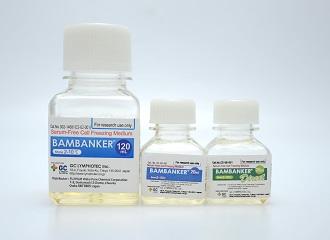
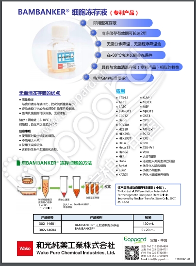
BAMBANKER® 是一种无血清细胞冻存液。可在 -80℃ 长期保存细胞(肿瘤细胞和常规细胞)。

BAMBANKER® 无血清细胞冻存液                              BAMBANKER®

◆产品特性

 ● 即用型细胞冻存液

 ● 无需分步降温,直接使用

 ● 无需稀释

 ● 无需程序降温盒

 ● -80℃ 长期保存

 ● 无血清

无血清冻存液的优点

 ● 与含血清类型相比,批次间的成分组成差异小,可保持稳定的品质。

 ● 不含血清,因此没有因动物源的未知成分和感染物质所产生的影响与风险。

 ● 可对无血清驯化细胞进行冷冻,节省再驯化步骤。


操作流程

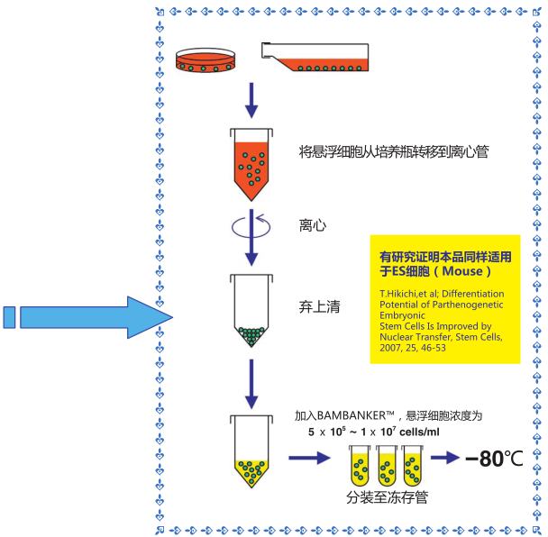
1)收集生长对数期*的细胞(5×10⁵-1×10⁷个细胞)

2)用1mL 该细胞冻存液悬浮细胞,置于冻存管中,不需预冷,直接 -80℃ 冷冻保存,也可-80℃冻存 12 小时后可

      转移至液氮中保存。

3)用恒温箱或者水浴锅快速复苏细胞

      *冷冻细胞必须处于生长对数期

BAMBANKER® 无血清细胞冻存液                              BAMBANKER®


无菌检测**

内毒素:生色底物法

支原体:荧光抗体法

真菌和细菌:依据日本药典

**:可索取检验证书)


BAMBANKER® Direct

BAMBANKER Direct 是“无血清型”细胞冻存液。

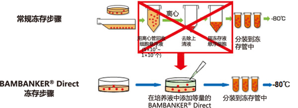
BAMBANKER® Direct无需离心收集细胞

BAMBANKER® 无血清细胞冻存液                              BAMBANKER®① 不需冻存前预处理,操作简便

② 不需稀释,直接使用

③ 无需分步降温,直接使用

④ 可快速、长期冻存细胞(-80℃或液氮)

⑤ 不含血清


BAMBANKER® 无血清细胞冻存液                              BAMBANKER®


BAMBANKER® Direct冻存步骤VS常规冻存步骤

使用本产品无需经过离心等复杂步骤,只需往培养基内添加与培养液等量的 BAMBANKER Direct,再分装到冻存管,置于-80℃ 便可冻存细胞。


应用

BAMBANKER使用例】

 细胞名称  保存时间
生存率
  BAMBANKER  公司A(含血清)
公司A(不含血清)

P3U1

(小鼠骨髓瘤细胞系)

12个月
95% 95% 70%

K562

(人白血病细胞系)

12个月
73% 70% 60%
人体胃黏膜上皮细胞 10个月
100% 62% 56%

human γδT cells

(人γδT细胞)

10个月
65% 37% 35%

 Daudi

(人 B细胞系)

12个月
100% 100% 92%

PC12

(大鼠源肾上腺嗜铬细胞瘤)

11个月
95% 59% 20%

human B cell line 

(人B细胞系)

9个月
74% 54% 35%

OKT4

(小鼠杂交瘤细胞)

12个月
100% 100% 92%

B细胞系

(猴)

10个月 56% 40% 18%

低温冻存实验证明以下细胞保存完好


3T3‐L1(小鼠前脂肪细胞系)

A431(人扁平上皮癌细胞系)

BAEC(牛主动脉血管内皮细胞系)

Balb/3T3(小鼠成纤维细胞系)

C2C12(小鼠骨骼肌细胞系)

 Daudi(人B细胞系)

ECV304(人脐静脉内皮细胞系)

H295R(肾上腺皮质细胞)

HEK293(人胚胎肾细胞系)

HEK293T(人胚胎肾细胞系)

HeLa(人子宫颈癌细胞系)

HeLa S3(人子宫颈癌细胞系)

HepG2(人肝癌细胞系)

HFF(人正常成纤维细胞系)

Huh7(人肝癌细胞系)

Jurkat(人白血病T细胞系)

K562(人慢性骨髓性白血病细胞系)

KATOIII (人胃癌上皮细胞系)

KLM‐1(人胰腺癌细胞系)

MDCK(犬肾小管上皮细胞系)

MEF(小鼠胚胎成纤维细胞)

NIH3T3(小鼠胚胎皮肤细胞)

OKT4(小鼠杂交瘤细胞)

OP9(小鼠骨髓基质细胞)

P3U1(小鼠骨髓瘤细胞系)

PANC‐1(人胰腺癌细胞系)

PC12(大鼠源肾上腺嗜铬细胞瘤)

RPE(人视网膜上皮细胞系)

SNL(小鼠胚胎成纤维细胞)

TSU‐Pr1(人前列腺癌细胞系)

Vero(非洲绿猴肾细胞系)

human γδT cells (人γδT细胞)

human B cell line (人B细胞系)

HDF(人皮肤成纤维细胞)  HCC20(人乳腺原发性导管癌细胞) BMMCs(人骨髓单核细胞系)
BMMCs(猪骨髓单核细胞系) BMSCs(马骨髓间充质干细胞系) C1(人成纤维细胞系)
CEF(牛胚胎成纤维细胞) CHO-K1(中国仓鼠卵巢细胞系) DPCs(大鼠牙髓细胞)
DPCs(人牙髓细胞) ESCs(人胚胎干细胞) EVT(人绒毛外滋养层细胞)
GH3(大鼠垂体瘤细胞) Gli36(胶质瘤细胞系)
h1(人类胚胎干细胞)
h9(人类胚胎干细胞) HN4(人口腔上皮细胞系)

HS-RMS-2

(多形性横纹肌肉瘤细胞系)

IPS(人诱导性多能干细胞)

LNCaP clone FGC

(人前列腺癌细胞)

MCF 10A(人正常乳腺细胞)

MEF-BL/6-1

(小鼠胚胎成纤维细胞)

MNCs(人单核细胞) MSCs(大鼠间充质干细胞系)
PBMCs(人外周血单个核细胞) PDL(人牙周膜细胞) pES(大鼠孤雌胚胎干细胞系)
Sf9(草地贪夜蛾细胞系) U251(胶质瘤细胞系) U87(胶质瘤细胞系)
VT(人绒毛膜滋养层细胞) 癌症干细胞 大鼠肝细胞

猴B细胞系

人外周血活化淋巴细胞

永生化人肌肉细胞

小鼠脾脏活化淋巴细胞

小鼠ES细胞系

人胃上皮细胞

大鼠神经祖细胞 大鼠脂肪细胞 狗肿瘤细胞
胶质瘤细胞系 牛脂肪细胞 牛子宫内膜上皮细胞
人扁桃体细胞 人肝细胞 人骨髓CD34+细胞
人巨噬细胞 人淋巴细胞 人输卵管上皮细胞
人胎儿卵巢成纤维细胞 人胎儿卵巢体细胞 人自然杀伤细胞
神经祖细胞 小鼠颅骨成骨细胞 心肌祖细胞
猪成纤维细胞


ES细胞(小鼠)使用实例
T.Hikichi,et al; Differentiation Potential of Parthenogenetic Embryonic Stem Cells Is Improved by Nuclear Transfer, Stem Cells, 2007, 25, 46-53

更多相关资料请点击文字:

BAMBANKER® 与自制冻存液的冻存效果比较


※ 本页面产品仅供研究用。研究以外不可使用。


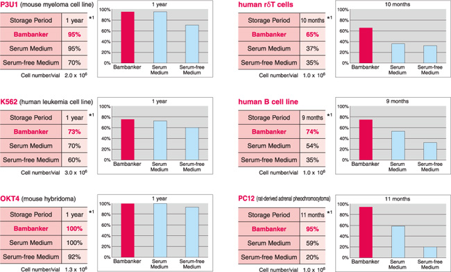
Bambanker® 与其他相关产品的比较

细胞冻存效果验证


细胞冻存液类型

1.Bambanker®

2.Medium with serum (含血清,A公司)

3.Serum-free Medium (无血清,A公司)


实验结果

*1:细胞-80℃的保存时间


BAMBANKER® 无血清细胞冻存液                              BAMBANKER®


相关PDF


BAMBANKER® 无血清细胞冻存液                              BAMBANKER®

BAMBANKER® 无血清细胞冻存液

BAMBANKER® 无血清细胞冻存液                              BAMBANKER®

Wako BAMBANKER冻存液(新手册)

细胞种类列举

BAMBANKER® 无血清细胞冻存液                              BAMBANKER®

BAMBANKER细胞冻存液

冻存解冻步骤说明

(终端).pdf

1.

Q:为什么我使用了 BAMBANKER® 来保存细胞,但是存活率依然不高?

A:请冻存前确保细胞处于生长对数期,并且冻存时细胞数目控制在 5×10⁵~1×10⁷/mL 冻存液。

2.

Q:我们实验室已经有固定的冻存程序了,换了你们的 BAMBANKER® 可以还继续用原来程序降温的方法冻存吗?

A:虽然本产品可以无需程序降温冻存细胞,但如果通过程序降温盒等适当控制了温度下降的速度,效果更佳。

3.

Q:哪些细胞株(系)适合使用BAMBANKER® 来进行细胞冷冻保存?

A:几乎所有细胞株(系)都可以使用 BAMBANKER® 进行冻存。对于较为宝贵的 ES/iPS 细胞的保存尤其适用。官网上所列举的细胞系均已经过测试验证。

但也并不排除可能有某些细胞株是不适合使用 BAMBANER® 来进行冻存的,用户在没有确认是否可使用时,建议在进行正式细胞冻存之前先进行预实验。

4.

Q:无血清冻存液相比传统含血清冻存液有什么优势?

A:无血清冻存液因不含有动物血清,质量更稳定,批间差小;同时未知生物成分或感染性物质污染细胞的几率也极低,尤其对于 ES/iPS 细胞等有可能用于再生医疗的细胞安全得以严格保障;可以直接冻存无血清培养的细胞,免去无血清再驯化的步骤;另外 BAMBANKER® 无血清细胞冻存液不需要像传统的血清冻存液需要程序降温,减少了用户的繁琐操作,节省了时间。

5.

Q:BAMBANKER® 在冷冻保存细胞的过程中起什么作用?

A:BAMBANKER® 无血清细胞冻存液使用了 DMSO 等作为保护剂,在冻存细胞时能以1℃/min 左右的温度下降而逐渐冻结,在此过程中,细胞内的水分子被置换成冻结保护剂,抑制胞内和细胞周边的冰晶的形成,防止细胞膜和细胞器结构损伤,防止蛋白变质。

6.

Q:未使用的BAMBANKER® 无血清细胞冻存液应该如何保存?

A:2-10℃ 避光保存。开封后尽快使用。请注意保质期为自生产日期起 24 个月。

7.

Q:BAMBANKER® 能否使用于医疗领域?

A:BAMBANKER® 仅供科研使用,不能使用于人体或医疗领域。

BAMBANKER™参考文献


[1]

Zhang C., Seo J., Nakamura T.   (2018) Cellular Approaches in Investigating Argonaute2-Dependent RNA   Silencing. In: Okamura K., Nakanishi K. (eds) Argonaute Proteins. Methods in   Molecular Biology, vol 1680. Humana Press, New York, NY.

[2]

Sharma, A., M¨ucke, M., &   Seidman, C. E. (2018). Human induced pluripotent stem cell production and   expansion from blood using a non-integrating viral reprogramming vector.   Current Protocols in Molecular Biology,122, e58. doi: 10.1002/cpmb.58.

[3]

Souta Motoike, Mikihito Kajiya,   Nao Komatsu, et al. Cryopreserved clumps of mesenchymal stem   cell/extracellular matrix complexes retain osteogenic capacity and induce   bone regeneration. Stem Cell Res Ther. 2018; 9: 73. Published online 2018 Mar   21. doi: 10.1186/s13287-018-0826-0.

[4]

Konuma T1, Kohara C1, Watanabe   E2, et al. Monocyte subsets and their phenotypes during treatment with   BCR-ABL1 tyrosine kinase inhibitors for Philadelphia chromosome-positive   leukemia. Hematol Oncol. 2018 Apr;36(2):451-456. doi: 10.1002/hon.2497. Epub   2018 Feb 12.

[5]

Srijaya Thekkeparambil   Chandrabose, Sandhya Sriram, et al. Amenable epigenetic traits of dental pulp   stem cells underlie high capability of xeno-free episomal reprogramming.

Stem Cell Research & Therapy 2018 9:68.

[6]

Evans, Michael A. et al.   "Macrophage-Mediated Delivery of Light Activated Nitric Oxide Prodrugs with   Spatial, Temporal and Concentration Control." Chemical Science (2018): n.   pag. Web. doi:10.1039/C8SC00015H.

[7]

Jauregui, C.; Yoganarasimha, S.;   Madurantakam, P. Mesenchymal Stem Cells Derived from Healthy and Diseased   Human Gingiva Support Osteogenesis on Electrospun Polycaprolactone Scaffolds.   Bioengineering 2018, 5, 8.

[8]

Khamaikawin, Wannisa et al.   Modeling Anti-HIV-1 HSPC-Based Gene Therapy in Humanized Mice Previously   Infected with HIV-1. Molecular Therapy – Methods & Clinical Development ,   Volume 9,23-32.

[9]

Masako Okumura, Toyoaki Natsume,   Masato T Kanemaki, Tomomi Kiyomitsu. Optogenetic reconstitution reveals that   Dynein-Dynactin-NuMA clusters generate cortical spindle-pulling forces as a   multi-arm ensemble. bioRxiv 277202; doi: https://doi.org/10.1101/277202

[10]

https://labchem.wako-chem.co.jp/journal/docs/proup10.pdf<链接>

[11]

Ince T A, Aster J C. In vitro   culture conditions for T-cell acute lymphoblastic leukemia/lymphoma: U.S.   Patent 9,683,217[P]. 2017-6-20.

[12]

Morris C D, Azadnia P, de Val N,   et al. Differential Antibody Responses to Conserved HIV-1 Neutralizing   Epitopes in the Context of Multivalent Scaffolds and Native-Like gp140   Trimers[J]. mBio, 2017, 8(1): e00036-17.<链接>

[13]

Lee K, Saetern O C, Nguyen A, et   al. Derivation of Leptomeninges Explant Cultures from Postmortem Human Brain   Donors[J]. JoVE (Journal of Visualized Experiments), 2017 (119):   e55045-e55045.<链接>

[14]

Buenrostro J D, Corces R, Wu B,   et al. Single-cell epigenomics maps the continuous regulatory landscape of   human hematopoietic differentiation[J]. bioRxiv, 2017: 109843.<链接>

[15]

Edmonds R E, Garvican E R, Smith   R K W, et al. Influence of commonly used pharmaceutical agents on equine bone   marrow‐derived mesenchymal stem cell viability[J]. Equine veterinary journal,   2017, 49(3): 352-357.<链接>

[16]

Jitraruch S, Dhawan A, Hughes R   D, et al. Cryopreservation of Hepatocyte Microbeads for Clinical   Transplantation[J]. Cell transplantation, 2017.<链接>

[17]

Usarek E, Barańczyk-Kuźma A,   Kaźmierczak B, et al. Validation of qPCR reference genes in lymphocytes from   patients with amyotrophic lateral sclerosis[J]. PloS one, 2017, 12(3):   e0174317.<链接>

[18]

Gagnon E, Connolly A, Dobbins J,   et al. Studying Dynamic Plasma Membrane Binding of TCR-CD3 Chains During   Immunological Synapse Formation Using Donor-Quenching FRET and FLIM-FRET[J].   The Immune Synapse: Methods and Protocols, 2017: 259-289.<链接>

[19]

Foster K, Chaddock J, Penn C, et   al. Non-cytotoxic protein conjugates: U.S. Patent 9,474,807[P]. 2016-10-25.

[20]

Araki N, Iida M, Machida K.   Bioassay method for detecting physiologically active substance: U.S. Patent   9,316,588[P]. 2016-4-19.

[21]

Sazinsky S, Michaelson J S,   Sathyanarayanan S, et al. Antibodies to icos: U.S. Patent Application   15/076,867[P]. 2016-3-22

[22]

李凯. Studies on Innate Immune   Activation by HBV Infection and Its Sensing Mechanism in Hepatocytes[J].   2016.

[23]

Ip L R H. Effect of INPP4B loss   on DNA repair and treatment strategies in ovarian cancer[D]. UCL (University   College London), 2016.

[24]

Thakkar A. Novel hormonal   combination therapy for triple negative breast cancer[D]. University of   Miami, 2016.

[25]

Pakdaman Y. In-vitro   characterization of STUB1 mutations in recessively inherited spinocerebellar   ataxia-16[D]. The University of Bergen, 2016.

[26]

Caxaria S. Induced pluripotent   stem cells (iPSCs) for research and therapy: induction of hepatic   differentiation in iPSCs and evaluation of their quality as a model of in   vivo development in the context of coagulation[D]. UCL (University College   London), 2016.<链接>

[27]

Bayne R A, Donnachie D J,   Kinnell H L, et al. BMP signalling in human fetal ovary somatic cells is   modulated in a gene-specific fashion by GREM1 and GREM2[J]. MHR: Basic   science of reproductive medicine, 2016, 22(9): 622-633.<链接>

[28]

Friedrich D. HIF-1 [alpha]   Drives Fungal Immunity in Human Macrophages[D]. Universität zu Lübeck,   2016.<链接>

[29]

Yasuda M, Kawabata J,   Akieda-Asai S, et al. Guanylyl cyclase C and guanylin reduce fat droplet   accumulation in cattle mesenteric adipose tissue[J]. The Journal of   Veterinary Science, 2016.<链接>

[30]

Campa M J, Moody M A, Zhang R,   et al. Interrogation of individual intratumoral B lymphocytes from lung   cancer patients for molecular target discovery[J]. Cancer Immunology,   Immunotherapy, 2016, 65(2): 171-180.<链接>

[31]

Kobayashi T, Yagi Y, Nakamura T.   Development of Genome Engineering Tools from Plant-Specific PPR Proteins   Using Animal Cultured Cells[J]. Chromosome and Genomic Engineering in Plants:   Methods and Protocols, 2016: 147-155.<链接>

[32]

Shikata H, Kaku M, Kojima S I,   et al. The effect of magnetic field during freezing and thawing of rat bone   marrow-derived mesenchymal stem cells[J]. Cryobiology, 2016, 73(1):   15-19.<链接>

[33]

Hirakawa M, Matos T, Liu H, et   al. Low-dose IL-2 selectively activates subsets of CD4+ Tregs and NK   cells[J]. JCI insight, 2016, 1(18).<链接>

[34]

Durruthy-Durruthy J, Sebastiano   V, Wossidlo M, et al. The primate-specific noncoding RNA HPAT5 regulates   pluripotency during human preimplantation development and nuclear   reprogramming[J]. Nature genetics, 2016, 48(1): 44-52.<链接>

[35]

Caxaria S, Arthold S, Nathwani A   C, et al. Generation of integration-free patient specific iPS cells using   episomal plasmids under feeder free conditions[J]. Patient-Specific Induced   Pluripotent Stem Cell Models: Generation and Characterization, 2016: 355-366.<链接>

[36]

Nonomura Y, Otsuka A, Nakashima   C, et al. Peripheral blood Th9 cells are a possible pharmacodynamic biomarker   of nivolumab treatment efficacy in metastatic melanoma patients[J].   Oncoimmunology, 2016, 5(12): e1248327.<链接>

[37]

Burridge P W, Diecke S, Matsa E,   et al. Modeling cardiovascular diseases with patient-specific human   pluripotent stem cell-derived cardiomyocytes[J]. Patient-Specific Induced   Pluripotent Stem Cell Models: Generation and Characterization, 2016: 119-130.<链接>

[38]

Mendonça M C P, Soares E S, de   Jesus M B, et al. PEGylation of Reduced Graphene Oxide Induces Toxicity in   Cells of the Blood–Brain Barrier: An in Vitro and in Vivo Study[J]. Molecular   pharmaceutics, 2016, 13(11): 3913-3924.<链接>

[39]

Eldaim A, Hashimoto O, Ohtsuki   H, et al. Expression of uncoupling protein 1 in bovine muscle cells[J].   Journal of animal science, 2016, 94(12): 5097-5104.<链接>

[40]

Zhen A, Rezek V, Youn C, et al.   Stem-cell based engineered immunity against HIV infection in the humanized   mouse model[J]. JoVE (Journal of Visualized Experiments), 2016 (113):   e54048-e54048.<链接>

[41]

Bastian N A, Bayne R A,   Hummitzsch K, et al. Regulation of fibrillins and modulators of TGFβ in fetal   bovine and human ovaries[J]. Reproduction, 2016, 152(2): 127-137.<链接>

[42]

Eto K, Takayama N, Nakamura S,   et al. Method for producing differentiated cells: U.S. Patent 9,200,254[P].   2015-12-1.

[43]

Eto K, Takayama N, Nakamura S,   et al. Novel Method for Producing Differentiated Cells: U.S. Patent   Application 14/925,508[P]. 2015-10-28.

[44]

Yamashita J, Takeda M.   Cd82-positive cardiac progenitor cells: U.S. Patent Application   15/308,147[P]. 2015-4-16.

[45]

Cai Y, Sugimoto C, Arainga M, et   al. Preferential Destruction of Interstitial Macrophages over Alveolar   Macrophages as a Cause of Pulmonary Disease in Simian Immunodeficiency   Virus–Infected Rhesus Macaques[J]. The Journal of Immunology, 2015, 195(10):   4884-4891.<链接>

[46]

Kojima S I, Kaku M, Kawata T, et   al. Cranial suture-like gap and bone regeneration after transplantation of   cryopreserved MSCs by use of a programmed freezer with magnetic field in   rats[J]. Cryobiology, 2015, 70(3): 262-268.<链接>

[47]

Egawa E Y, Kitamura N, Nakai R,   et al. A DNA hybridization system for labeling of neural stem cells with SPIO   nanoparticles for MRI monitoring post-transplantation[J]. Biomaterials, 2015,   54: 158-167.<链接>

[48]

Durruthy J D, Sebastiano V.   Derivation of GMP-Compliant Integration-Free hiPSCs Using Modified mRNAs[J].   Stem Cells and Good Manufacturing Practices: Methods, Protocols, and   Regulations, 2015: 31-42.<链接>

[49]

Sato Y, Sasaki T, Takahashi S,   et al. Development of a highly reproducible system to evaluate inhibition of   cytochrome P450 3A4 activity by natural medicines[J]. Journal of Pharmacy   & Pharmaceutical Sciences, 2015, 18(4): 316-327.<链接>

[50]

Burridge P W, Holmström A, Wu J   C. Chemically defined culture and cardiomyocyte differentiation of human   pluripotent stem cells[J]. Current protocols in human genetics, 2015: 21.3.   1-21.3. 15.<链接>

[51]

Käding N. Hypoxia Regulates Host   Cell Metabolism and Thereby Enhancing Clamydia Pneumonia Growth[D]. Zentrale   Hochschulbibliothek Lübeck, 2015.<链接>

[52]

Lu S. Calcium Dependent   Regulatory Mechanism in Wolfram Syndrome: A Dissertation[J]. 2015.

[53]

Garvican E R, Cree S, Bull L, et   al. Viability of equine mesenchymal stem cells during transport and   implantation[J]. Stem cell research & therapy, 2014, 5(4): 1.<链接>

[54]

Deng X, Terunuma H, Nieda M.   Method for producing nk cell-enriched blood preparation: U.S. Patent   Application 14/508,745[P]. 2014-10-7.

[55]

Foster K, Chaddock J, Penn C, et   al. Non-cytotoxic protein conjugates: U.S. Patent 8,778,634[P]. 2014-7-15.

[56]

Ramathal C Y, Dumuthy-Durruthy   J, Pera R A R, et al. Generation of male germ cells: U.S. Patent Application   14/904,396[P]. 2014-7-10

[57]

Ince T A. Assays, methods and   kits for analyzing sensitivity and resistance to anti-cancer drugs,   predicting a cancer patient's prognosis, and personalized treatment   strategies: U.S. Patent Application 14/894,595[P]. 2014-6-4.

[58]

Nishio M, Saeki K.   Differentiation of human pluripotent stem cells into highly functional   classical brown adipocytes[J]. Methods Enzymol, 2014, 537: 177-197.<链接>

[59]

Durruthy-Durruthy J, Briggs S F,   Awe J, et al. Rapid and efficient conversion of integration-free human   induced pluripotent stem cells to GMP-grade culture conditions[J]. PloS one,   2014, 9(4): e94231.<链接>

[60]

Koido S, Homma S, Okamoto M, et   al. Treatment with Chemotherapy and Dendritic Cells Pulsed with Multiple   Wilms' Tumor 1 (WT1)–Specific MHC Class I/II–Restricted Epitopes for   Pancreatic Cancer[J]. Clinical Cancer Research, 2014, 20(16):   4228-4239.<链接>

[61]

Patz Jr E F. Antibodies   Expressed by Intratumoral B Cells as the Basis for a Diagnostic Test for Lung   Cancer[R]. DUKE UNIV DURHAM NC, 2014.<链接>

[62]

Kaku M, Shimasue H, Ohtani J, et   al. A case of tooth autotransplantation after long-term cryopreservation   using a programmed freezer with a magnetic field[J]. The Angle Orthodontist,   2014, 85(3): 518-524.<链接>

[63]

Kaku M, Koseki H, Kojima S, et   al. Cranial bone regeneration after cranioplasty using cryopreserved   autogenous bone by a programmed freezer with a magnetic field in rats[J].   CryoLetters, 2014, 35(6): 451-461.<链接>

[64]

Koido S, Kinoshita S, Mogami T,   et al. Immunological assessment of cryotherapy in breast cancer patients[J].   Anticancer research, 2014, 34(9): 4869-4876.<链接>

[65]

Sazuka S, Katsuno T, Nakagawa T,   et al. Fibrocytes are involved in inflammation as well as fibrosis in the   pathogenesis of Crohn's disease[J]. Digestive diseases and sciences, 2014,   59(4): 760-768.<链接>

[66]

Lin S L, Lee S Y, Lin Y C, et   al. Evaluation of mechanical and histological properties of cryopreserved   human premolars under short-term preservation: A preliminary study[J].   Journal of Dental Sciences, 2014, 9(3): 244-248.<链接>

[67]

Poole E, Reeves M, Sinclair J H.   The use of primary human cells (fibroblasts, monocytes, and others) to assess   human cytomegalovirus function[J]. Human Cytomegaloviruses: Methods and   Protocols, 2014: 81-98.<链接>

[68]

Garvican E R, Dudhia J, Alves A   L, et al. Mesenchymal stem cells modulate release of matrix proteins from   tendon surfaces in vitro: a potential beneficial therapeutic effect[J].   Regenerative medicine, 2014, 9(3): 295-308.<链接>

[69]

Skinner J A, Zurawski S M,   Sugimoto C, et al. Immunologic characterization of a rhesus macaque H1N1   challenge model for candidate influenza vaccine assessment[J]. Clinical and   Vaccine Immunology, 2014: CVI. 00547-14.<链接>

[70]

Terunuma H, Deng X, Nieda M.   Method for producing nk cell-enriched blood preparation: U.S. Patent   Application 14/780,394[P]. 2013-3-27.

[71]

Cho M, Yamazaki T, Endo M, et   al. Anti-Phospholipase D4 Antibody: U.S. Patent Application 14/375,266[P].   2013-1-31.

[72]

Bhandari S. Radiological,   clinical and laboratory based studies in the pathogenesis of desmoid tumours   in familial adenomatous polyposis[J]. 2013.

[73]

Gonzàlez Juncà A. Study of   molecular mechanisms implicated in the TGF-beta oncogenic effect in   Glioma[J]. 2013.

[74]

Koseki H, Kaku M, Kawata T, et   al. Cryopreservation of osteoblasts by use of a programmed freezer with a   magnetic field[J]. CryoLetters, 2013, 34(1): 10-19.<链接>

[75]

Naito H, Yoshimura M, Mizuno T,   et al. The advantages of three‐dimensional culture in a collagen hydrogel for   stem cell differentiation[J]. Journal of Biomedical Materials Research Part   A, 2013, 101(10): 2838-2845.<链接>

[76]

Stec M, Baran J, Szatanek R, et   al. Properties of monocytes generated from haematopoietic CD34+ stem cells   from bone marrow of colon cancer patients[J]. Cancer Immunology,   Immunotherapy, 2013, 62(4): 705-713.<链接>

[77]

Müller L, Brighton L E, Carson J   L, et al. Culturing of human nasal epithelial cells at the air liquid   interface[J]. Journal of visualized experiments: JoVE, 2013 (80).<链接>

[78]

Kalaszczynska I, Ruminski S,   Platek A E, et al. Substantial differences between human and ovine   mesenchymal stem cells in response to osteogenic media: how to explain and   how to manage?[J]. BioResearch open access, 2013, 2(5): 356-363.<链接>

[79]

Tamai Y, Hasegawa A, Takamori A,   et al. Potential Contribution of a Novel Tax Epitope–Specific CD4+ T Cells to   Graft-versus-Tax Effect in Adult T Cell Leukemia Patients after Allogeneic   Hematopoietic Stem Cell Transplantation[J]. The Journal of Immunology, 2013,   190(8): 4382-4392.<链接>

[80]

Kasai K, Nakashima H, Liu F, et   al. Toxicology and biodistribution studies for MGH2. 1, an oncolytic virus   that expresses two prodrug-activating genes, in combination with prodrugs[J].   Molecular Therapy-Nucleic Acids, 2013, 2: e113.<链接>

[81]

Deng X, Terunuma H, Nieda M.   Method for producing nk cell-enriched blood preparation: U.S. Patent   Application 13/980,777[P]. 2012-1-17.

[82]

Somm E, Bonnet N, Martinez A, et   al. A botulinum toxin–derived targeted secretion inhibitor downregulates the   GH/IGF1 axis[J]. The Journal of clinical investigation, 2012, 122(9):   3295.<链接>

[83]

Takaoka E, Sonobe H, Akimaru K,   et al. Multiple sites of highly amplified DNA sequences detected by molecular   cytogenetic analysis in HS-RMS-2, a new pleomorphic rhabdomyosarcoma cell   line[J]. American journal of cancer research, 2012, 2(2): 141.<链接>

[84]

Fahlbusch F B, Dawood Y, Hartner   A, et al. Cullin 7 and Fbxw 8 expression in trophoblastic cells is regulated   via oxygen tension: implications for intrauterine growth restriction?[J]. The   Journal of Maternal-Fetal & Neonatal Medicine, 2012, 25(11): 2209-2215.<链接>

[85]

Aloé S, Weber F, Behr B, et al.   Modulatory effects of bovine seminal plasma on uterine inflammatory   processes[J]. Reproduction in domestic animals, 2012, 47(1): 12-19.<链接>

[86]

Gupta A, Bhakta S. An integrated   surrogate model for screening of drugs against Mycobacterium tuberculosis[J].   Journal of antimicrobial chemotherapy, 2012, 67(6): 1380-1391.<链接>

[87]

Saeki K. Feeder-Free Culture for   High Efficiency Production of Subculturable Vascular Endothelial Cells from   Human Embryonic Stem Cells[J]. Human Embryonic and Induced Pluripotent Stem   Cells: Lineage-Specific Differentiation Protocols, 2012: 277-294.<链接>

[88]

Yamazaki T, Okabe H, Kobayashi   S, et al. Cancer stem cell mass and process for production thereof: U.S.   Patent Application 13/878,181[P]. 2011-10-6.

[89]

Deng X, Terunuma H, Nieda M.   Method for producing nk cell-enriched blood product: U.S. Patent Application   13/577,476[P]. 2011-2-4.

[90]

Sugii S, Kida Y, Berggren W T,   et al. Feeder-independent ips cell derivation from human and mouse adipose   stem cells[J]. Nature protocols, 2011, 6(3): 346.<链接>

[91]

Shinada T, Akimoto T, Zhu Y, et   al. Modulation of viability of live cells by focused ion‐beam exposure[J].   Biotechnology and bioengineering, 2011, 108(1): 222-225.<链接>

[92]

Huang M S, Chang W J, Huang H M,   et al. Effects of transportation time after extraction on the magnetic   cryopreservation of pulp cells of rat dental pulp[J]. Journal of Dental   Sciences, 2011, 6(1): 48-52.<链接>

[93]

Sato D, Suzuki Y, Kano T, et al.   Tonsillar TLR9 expression and efficacy of tonsillectomy with steroid pulse   therapy in IgA nephropathy patients[J]. Nephrology Dialysis Transplantation,   2011, 27(3): 1090-1097.<链接>

[94]

Kamada H, Kaku M, Kawata T, et   al. In-vitro and in-vivo study of periodontal ligament cryopreserved with a   magnetic field[J]. American Journal of Orthodontics and Dentofacial   Orthopedics, 2011, 140(6): 799-805.<链接>

[95]

Bui H T, Wakayama S, Mizutani E,   et al. Essential role of paternal chromatin in the regulation of   transcriptional activity during mouse preimplantation development[J].   Reproduction, 2011, 141(1): 67-77.<链接>

[96]

Takata Y, Kishine H, Sone T, et   al. Generation of iPS cells using a BacMam multigene expression system[J].   Cell structure and function, 2011, 36(2): 209-222.<链接>

[97]

Benko Z, Zhao R Y. Zeocin for   selection of bleMX6 resistance in fission yeast[J]. Biotechniques, 2011,   51(1): 57-60.<链接>

[98]

Abedini S, Kaku M, Kawata T, et   al. Effects of cryopreservation with a newly-developed magnetic field   programmed freezer on periodontal ligament cells and pulp tissues[J].   Cryobiology, 2011, 62(3): 181-187.<链接>

[99]

Oshima-Sudo N, Li Q, Hoshino Y,   et al. Optimized method for culturing outgrowth endothelial progenitor   cells[J]. Inflammation and Regeneration, 2011, 31(2): 219-227.<链接>

[100]

Araki N. Bioassay method for   antibody against thyroid-stimulating hormone receptor, measurement kit for   the antibody, and novel genetically modified cell for use in the bioassay   method or the measurement kit: U.S. Patent Application 13/381,402[P]. 2010-6-24.

[101]

Foster K, Chaddock J, Marks P,   et al. Fusion proteins: U.S. Patent 7,659,092[P]. 2010-2-9.

[102]

Mieno S, Boodhwani M, Robich M   P, et al. Effects of diabetes mellitus on VEGF‐induced proliferation response   in bone marrow derived endothelial progenitor cells[J]. Journal of cardiac   surgery, 2010, 25(5): 618-625.<链接>

[103]

Kaku M, Kamada H, Kawata T, et   al. Cryopreservation of periodontal ligament cells with magnetic field for   tooth banking[J]. Cryobiology, 2010, 61(1): 73-78.<链接>

[104]

Kawata T, Kaku M, Fujita T, et   al. Water molecule movement by a magnetic field in freezing for tooth   banking[J]. Biomedical Research, 2010, 21(4).<链接>

[105]

Lee S Y, Chiang P C, Tsai Y H,   et al. Effects of cryopreservation of intact teeth on the isolated dental   pulp stem cells[J]. Journal of Endodontics, 2010, 36(8): 1336-1340.<链接>

[106]

Huang Y H, Yang J C, Wang C W,   et al. Dental stem cells and tooth banking for regenerative medicine[J].   Journal of Experimental & Clinical Medicine, 2010, 2(3):   111-117.<链接>

[107]

Kwon H J, Enomoto T, Shimogawara   M, et al. Benchmarks[J]. Biotechniques, 2010, 48: 460-462.<链接>

[108]

Shimizu Y, Takamori A,   Utsunomiya A, et al. Impaired Tax‐specific T‐cell responses with insufficient   control of HTLV‐1 in a subgroup of individuals at asymptomatic and smoldering   stages[J]. Cancer science, 2009, 100(3): 481-489.<链接>

[109]

Park H S, Cho S G, Park M J, et   al. Bone marrow T cells are superior to splenic T cells to induce chimeric   conversion after non-myeloablative bone marrow transplantation[J]. The Korean   journal of internal medicine, 2009, 24(3): 252.<链接>

[110]

Enosawa S, Miyamoto Y, Ikeya T.   Frozen cell immobilized product, primary hepatocyte culture tool, and method   for producing primary hepatocyte culture tool: U.S. Patent Application   12/738,809[P]. 2008-9-11.

[111]

DePinho R A, Stommel J M.   Receptor tyrosine kinase profiling: U.S. Patent Application 12/450,820[P].   2008-4-11.

[112]

Mieno S, Clements R T, Boodhwani   M, et al. Characteristics and Function of Cryopreserved Bone Marrow–Derived   Endothelial Progenitor Cells[J]. The Annals of thoracic surgery, 2008, 85(4):   1361-1366.<链接>

[113]

Warren C. The Response of HN4   Cells to Porphyromonas gingivalis DNA[D]. , 2008.

[114]

Hikichi T, Wakayama S, Mizutani   E, et al. Differentiation potential of parthenogenetic embryonic stem cells   is improved by nuclear transfer[J]. Stem Cells, 2007, 25(1): 46-53.<链接>

[115]

Zaidi S K, Pande S, Pratap J, et   al. Runx2 deficiency and defective subnuclear targeting bypass senescence to   promote immortalization and tumorigenic potential[J]. Proceedings of the   National Academy of Sciences, 2007, 104(50): 19861-19866.<链接>

[116]

Hikichi T, Wakayama S, Mizutani   E, et al. Differentiation potential of parthenogenetic embryonic stem cells   is improved by nuclear transfer[J]. Stem Cells, 2007, 25(1): 46-53.<链接>

[117]

Liu D G, Kobayashi T, Onishi A,   et al. Relation between human decay‐accelerating factor (hDAF) expression in   pig cells and inhibition of human serum anti‐pig cytotoxicity: value of   highly expressed hDAF for xenotransplantation[J]. Xenotransplantation, 2007,   14(1): 67-73.<链接>

[118]

Ishii H, Iinuma A, Osumi K, et   al. Canine tumor treatment method, pharmaceutical formulation applied   thereto, and method of cryogenically preserving cells used therewith: U.S.   Patent Application 11/465,892[P]. 2006-8-21.

[119]

Hatoya S, Sugiyama Y, Torii R,   et al. Effect of co-culturing with embryonic fibroblasts on IVM, IVF and IVC   of canine oocytes[J]. Theriogenology, 2006, 66(5): 1083-1090.<链接>

[120]

Sasaki M, Kato Y, Yamada H, et   al. Development of a novel serum‐free freezing medium for mammalian cells   using the silk protein sericin[J]. Biotechnology and applied biochemistry,   2005, 42(2): 183-188.<链接>

[121]

Haynes J E. Pseudonyms of   Authors: Including Anonyms and Initialisms[M]. JE Haynes, 1882.<链接>

产品编号 产品名称 产品规格 产品等级
302-14681 BAMBANKER
 BAMBANKER冻存液
120 mL
306-14684 BAMBANKER
 BAMBANKER冻存液
20 mLx5
306-95921 BAMBANKER Direct
 BAMBANKER直接冻存液
20 mL

胰蛋白酶EDTA溶液(无酚红), AF Trypsin-EDTA Solution without Phenol Red, AF

  • 产品特性
  • 相关资料
  • Q&A
  • 参考文献

无动物源胰蛋白酶EDTA胰蛋白酶EDTA溶液(无酚红), AF                              Trypsin-EDTA Solution without Phenol Red, AF


  本品是已经过病原体、内毒素和无菌检测的胰蛋白酶EDTA溶液,可用于贴壁细胞的剥离、各组织的细胞分散等。

  本品以重组胰蛋白酶为原料,不含动物源物质,无需担心病毒污染,能让您更安心地进行实验。使用方法与0.05%胰蛋白酶-EDTA溶液一样。


◆特点:


 ● 不含动物源物质

 ● 高细胞剥离率,剥离后细胞生存率高


●产品规格项目:

规格

外观

pH

渗透压

无菌试验

内毒素测试

支原体检查

应用试验
  (Vero
细胞在10分钟后完全剥离,高达90%以上的细胞存活率)


●细胞剥离能力:


胰蛋白酶EDTA溶液(无酚红), AF                              Trypsin-EDTA Solution without Phenol Red, AF

[培养条件]

细胞:MDCK细胞

培养基:E-MEM+10%FBS

接种细胞数 : 5.0×10⁴cells/cm2

培养方法 :静置培养

培养器材: 12孔板

培养环境 : 37℃, 5% CO2, 4天

培养后、37℃, 5% CO2条件下处理15分钟,测定剥离细胞数。

→与其他产品相比有更好的细胞剥离率。

●继代培养时的影响:


胰蛋白酶EDTA溶液(无酚红), AF                              Trypsin-EDTA Solution without Phenol Red, AF

[培养条件]

细胞:MDCK细胞

培养基 : E-MEM+10% FBS

接种细胞数 : 3.0×10⁵cells

培养方法 : 静置培养

培养器材 : T25瓶

培养环境 : 37℃, 5% CO2

37℃, 5% CO2条件下进行15分钟胰蛋白酶处理后,进行继代培养

→细胞增殖不受本品影响。

查看相关产品请看相关资料


◆ 相关产品:


● 胰蛋白酶EDTA(使用猪细小病毒原料)


产品编号.

产品名称

规格

包装

202-16931

0.05w/v% Trypsin-0.53mmol/l EDTA・4Na Solution

with Phenol Red

0.05w/v% 胰蛋白酶-0.53mmol/l EDTA . 4Na溶液和酚红

细胞培养用

100 mL

204-16935

500 mL

209-16941

0.25w/v% Trypsin-1mmol/l EDTA・4Na Solution

with Phenol Red

100 mL

201-16945

500 mL

206-17291

0.5w/v% Trypsin-5.3mmol/l EDTA・4Na Solution

with Phenol Red (×10)

100 mL

208-17251

0.5w/v% Trypsin-5.3mmol/l EDTA・4Na Solution

without Phenol Red (×10)

100 mL


重组胰蛋白酶

生产商

产品编号

生产编号

产品名称

包装

Roche Diagnostics

罗氏诊断

631-24973

06369880103

Trypsin,Porcine,recombinant(Pichia pastoris),GMP Grade

1 g

635-24971

03358658103

3.5 MU

产品编号 产品名称 产品规格 产品等级
203-20251 Trypsin-EDTA Solution without Phenol Red, AF 100ml 细胞培养用
207-20271 Trypsin-EDTA Solution(High Trypsin) without Phenol Red, AF* 100ml 细胞培养用

细胞分散液 Accumax

  • 产品特性
  • 相关资料
  • Q&A
  • 参考文献


Accumax细胞分散液 Accumax

温和高效的细胞分散液

  Accumax 是天然酶混合物,用于组织分离,细胞计数以及分散细胞团块。

  Accumax 可直接代替胰蛋白酶和胶原酶。


特点


  ● 含水解蛋白酶和胶原酶的天然活性酶混合物

  ● 温和迅速

  ● 分散组织

  ● 提高手动或自动细胞计数的准确性
  ● 可用于分散神经细胞和前列腺球体 .

  ● 移除 3D 矩阵细胞

  ● 去除中空纤维细胞反应器中的细胞

  ● 减少流式细胞仪分选块状细胞的时间

  ● 无动物源,无细菌源成分

  ● 即用型(Dulbecco's PBS 预处理).

  ● 传代贴壁细胞无需中和

◆应用 案例


<已使用 Accumax 测试细胞系>


  人类胚胎干细胞,成纤维细胞,角化细胞,血管内皮细胞,肝细胞

  血管平滑肌细胞,肝细胞祖细胞,原代鸡胚神经细胞

  骨髓干细胞,粘附 CHO 和 BHK 细胞,巨噬细胞,293细胞,L929细胞,

  永生小鼠睾丸生殖细胞,3T3,Vero 细胞,COS,HeLa 细胞,NT2,MG63,M24 和 A375

  转移性黑素瘤,神经胶质瘤 U251 和 D54,HT1080 纤维肉瘤细胞和 Sf9 昆虫细胞.


<AccumaxAccutase 的区别>

产品

Accumax

Accutase

酶活性

正常

反应停止/中和

不需要

动物和细菌成分

不包含

酚红

不包含

包含


<Accumax 的数据

细胞分散液 Accumax


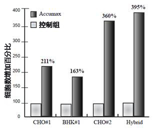
细胞等分后用等体积的 PBS(对照)或 Accumax 处理,并在37℃孵育5分钟。然后用细胞计数器机计数。

使用 Accumax 可有效将结块的细胞进行分散,获得更好的单一细胞。


<提高细胞计数重复性的实验报告概述>


  1、获取结块细胞的代表性样品进行计数,取 0.5 或 1.0ml 置于12×75 mm管。

  2、加等体积 Accumax 至细胞样品,并在室温下孵育5到 10 分钟。

  3、按正常程序计数细胞。 注意细胞已被稀释了2倍。

产品名称

规格

编号

存储

制造商

价格/详细信息

Accumax

100 mL

AM105

-20℃

FIC

请联系

500 mL

AM105-500

-20℃

FIC

请联系


  注意:所有产品仅供研究使用。不用于诊断。

产品编号 产品名称 产品规格 产品等级
AM105 Accumax 100 mL
AM105-500 Accumax 500 mL

玻璃粘连蛋白(20-398aa),人,重组体,溶液

  • 产品特性
  • 相关资料
  • Q&A
  • 参考文献

玻璃粘连蛋白(20-398aa),人,重组体,溶液玻璃粘连蛋白(20-398aa),人,重组体,溶液

细胞粘附蛋白


  玻璃粘连蛋白由478个氨基酸配列组成,是存在于血清和细胞外基质的糖蛋白。它与纤连蛋白、层连蛋白统称为细胞粘附蛋白。玻璃粘连蛋白除具有细胞粘附作用和伸展作用,还与补体系统、凝血系统相关的多功能蛋白质。

  本品是重组蛋白,除了信号域,由 20-398 氨基酸片段构成。可作为研究组织修复·再生等技能及细胞培养的基础。


◆参考数据:


玻璃粘连蛋白(20-398aa),人,重组体,溶液

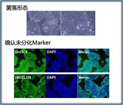
  在涂层6孔板用本品培养对hiPS细胞做6个继代培养,通过未分化 Marker,确认hiPS细胞未分化性的维持。
  细胞:hiPS201B7株
  培养基:StemSure® hPSC MediumΔ(产品编号197-17571)+ 32 ng/mL bFGF(产品编号060-05383) 


◆产品概要


  含量: 90%以上(SDS-PAGE)
  表达: E. coli
  状态: 溶液
  浓度: 0.5 mg/mL
  组份: 20 mM Tris-HCl,pH 8.0(含 NaCl,KCl,EDTA,精氨酸,DTT和甘油)


相关产品

  不含动物来源的无血清培养基,可进行无饲养层细胞培养人多功能干细胞,StemSure® hPSC培养基Δ。


产品编号

品名

规格

容量

197-17571

StemSure® hPSC培养基 Δ

细胞培养用

100 mL

193-17573

100 mL×4


产品编号

品名

规格

容量

029-18061

BC2LCN Lectin, recombinant, Solution【AilecS1】

糖链研究用

1 mg

025-18063

1 mg×5

064-05381

bFGF, Human, recombinant, Animal-derived-free

细胞生物学用

50 μg

068-05384

100 μg

060-05383

1 mg

180-02991

rBC2LCN-FITC 【AilecS1-FITC】 
  未分化细胞检测用

细胞染色用

100 μL

186-02993

100 μL×5

257-00511

Y-27632

细胞生物学用

1 mg

253-00513

5 mg

251-00514

25 mg

253-00591

5 nmol/l Y-27632 Solution

细胞培养用

300 μL


产品编号 产品名称 产品规格 产品等级
220-02041 Vitronectin(20-398 aa), Human, recombinant, Solution 500μg 生化学用

Culture® A-83-01

  • 产品特性
  • 相关资料
  • Q&A
  • 参考文献

Culture® A-83-01Culture® A-83-01

用于ES·IPS细胞研究


  本产品是ALK4、ALK5、ALK7的选择性抑制剂。可以抑制Smad2/3的磷酸化和TGF-β诱导性的上披肩组织转换。本产品对骨形成因子typeI受体、p38MAP激酶、细胞外抑制激酶几乎没有影响。

  另外,在大鼠IPS细胞培养基中添加本产品,可以抑制大鼠IPS细胞分化,进行均一增殖,从而可以长期培养。


Culture® A-83-01                            Culture® A-83-01


◆优点 

● 已确认细胞毒性

● 纯度(HPLC):98.0以上

● 外观:白色~黄色、结晶型粉末或块状

● 溶解性:可溶于DMSO


具体相关产品请查阅相关资料

欲了解更多相关产品请点击文字:ES/iPS细胞未分化维持研究用低分子化合物

相关产品

◆维持ES·IPS细胞的未分化能


产品编号

产品名称

规格

包装

013-22211

Adrenocorticotropic   Hormone

(1-24)(Human)【ACTH】

细胞生物学用

1mg

012-23021

ALK5 Inhibitor

【TGF-βRⅠ Kinase InhibitorⅡ】

细胞生物学用

1mg

021-17041

027-17043

(-)-Blebbistatin

细胞生物学用

1mg

5mg

029-16241

6-Bromoindirubin-3'-oxime【BIO】

【GSK-3 InhibitorⅨ】

细胞生物学用

1mg

029-05393

Butyric Acid

和光特级

25mL

023-05396

500mL

036-24001

Cyclic   Pifithrin-α Hydrobromide

细胞生物学用

5mg

041-30101

DNA   Methyltransferase Inhibitor

【RG108】

基因研究用

10mg

047-30103

25mg

056-08221

EHNA   Hydrochloride

细胞生物学用

10mg

079-03811

GF 109203X

生物化学用

1mg

086-10071

HA-100   Hydrochloride

细胞生物学用

10mg

110-00831

Kenpaullone

细胞生物学用

1mg

116-00833

5mg

115-01001

Ki16425

细胞生物学用

5mg

162-25291

PD0325901

细胞生物学用

5mg

168-25293

25mg

160-26831

PD173074

细胞生物学用

5mg

165-26761

PD184352

细胞生物学用

5mg

169-19211

PD-98059

生物化学用

5mg

198-16761

SB203580   Hydrochloride

细胞生物学用

1mg

193-01522

Sodium Butyrate

25g

197-01525

500g

193-16071

SU5402

细胞生物学用

1mg

202-18011

Thiazovivin

细胞生物学用

1mg

208-18013

5mg

211-01051

U0126

生物化学用

5mg

227-01071

Valproic Acid

生物化学用

5g

225-01072

25g

257-00511

Y-27632

细胞生物学用

1mg

253-00513

5mg

251-00514

25mg

253-00591

5mmol/ℓ Y-27632   Solution

细胞培养用

300μL


ES·IPS细胞的分化诱导、脱分化


产品编号

产品名称

规格

包装

011-25291

A-769662

细胞生物学用

5mg

017-25293

25mg

015-22531

AICAR

细胞生物学用

100mg

011-22533

1g

014-16631

Am580

生物化学用

5mg

014-25421

AMD3100   Octahydrochloride

细胞生物学用

10mg

031-18963

Ciclosporin A

细胞生物学用

50mg

035-18961

200mg

030-20981

Ciglitazone

细胞生物学用

5mg

034-21501

CKI-7   Dihydrochloride

细胞生物学用

5mg

043-33581

DAPT【γ-Secretase InhibitorⅨ】

细胞生物学用

5mg

049-33583

25mg

047-18863

Dexamethasone

生物化学用

100mg

041-18861

1g

067-02191

Forskolin

生物化学用

10mg

063-02193

25mg

092-07041

IPA-3

细胞生物学用

5mg

094-06381

IWR-1-endo

细胞生物学用

5mg

124-06011

LDN193189 Hydrochloride

细胞生物学用

2mg

129-04861

LY294002

生物化学用

5mg

125-04863

10mg

123-04864

25mg

166-23991

Purmorphamine

细胞生物学用

5mg

186-01114

all-trans-Retinoic   Acid

生物化学用

50mg

182-01116

100mg

182-01111

250mg

188-01113

1g

192-16541

SB431542

细胞生物学用

5mg

198-16543

25mg

198-09811

Spermine

生物化学用

250mg

194-09813

1g

203-17561

Trichostatin A

细胞生物学用

1mg

209-17563

5mg

209-19481

Troglitazone

细胞生物学用

5mg

205-19483

50mg


参考文献

[1]Li, W. et al. : Cell Stem Cell, 4, 16 2009.

产品编号 产品名称 产品规格 产品等级
039-24111 CultureSure ® A-83-01 2 mg 细胞培养用
035-24113 CultureSure ® A-83-01 10 mg 细胞培养用
细胞增殖因子

  • 产品特性
  • 相关资料
  • Q&A
  • 参考文献

细胞培养用培养基添加物细胞增殖因子

细胞増殖因子


  细胞增殖的相关物质。参与各种各样的细胞生物学、生理学过程的调节,通过与靶细胞表面受容体蛋白质的特异性结合,参与细胞间的信号传递。产品取自牛、人,植物重组体、大洋洲产地原料。


白蛋白


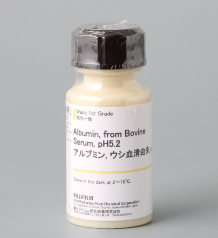
  白蛋白是可用于酵素和成长因子等的生物学性敏感型高分子稀释·稳定剂,或可添加到脂质培养基中难溶于水的成分。


细胞增殖因子


(产品编号:011-21271


◆胰岛素


  胰岛素是对生物体内的血糖调节、促进蛋白质合成有着重要作用的激素。可作为细胞培养中的增殖因子,促进蛋白质的合成。


细胞增殖因子


(产品编号:093-06471


转铁蛋白(Transferrin)


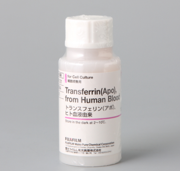
  转铁蛋白主要负责运转培养基中所含的铁离子。


细胞增殖因子

(产品编号:205-18121

 相关产品


细胞培养用产品 
 
  我司为您充分准备了液体培养基如平衡盐溶液、细胞剥离·分散用溶液、抗生物质溶液、细胞外基质等产品。

神经干细胞用添加剂


  作为血清替代品用于神经干细胞的培养。我司现有两款产品,一是用 Transferrin(Apo)调制而成的,一是用 Transferrin(Holo)调制而成的。一般产品都含 Transferrin(Holo),但是使用含有 Transferrin(Apo)的 N2 Supplement 的话,根据细胞类型的不同神经干细胞的增殖能力可不同层次的增加。

产品编号

品名

规格

容量

141-09041

N2 Supplement with Transferrin(Apo)(x 100)

N2神经细胞生长添加剂含转铁蛋白(Apo)(x 100)

细胞培养用

5 mL

141-08941

N2 Supplement with Transferrin(Holo)(x 100)

N2神经细胞生长添加剂含转铁蛋白(Holo)(x 100)

细胞培养用

5 mL


产品编号 产品名称 产品规格 产品等级
白蛋白
011-21271 Albumin, from Bovine Serum (BSA), pH5.2(Fraction V)牛血清白蛋白,pH5.2 1g 和光一级
017-21273 Albumin, from Bovine Serum (BSA), pH5.2(Fraction V)牛血清白蛋白,pH5.2 10g 和光一级
019-21272 Albumin, from Bovine Serum (BSA), pH5.2(Fraction V)牛血清白蛋白,pH5.2 25g 和光一级
015-21274 Albumin, from Bovine Serum (BSA), pH5.2(Fraction V)牛血清白蛋白,pH5.2 100g 和光一级
013-21275 Albumin, from Bovine Serum (BSA), pH5.2(Fraction V)牛血清白蛋白,pH5.2 500g 和光一级
011-21276 Albumin, from Bovine Serum (BSA), pH5.2(Fraction V)牛血清白蛋白,pH5.2 1kg 和光一级
013-23291 Albumin, from Bovine Serum (BSA), Cohn Fraction V, pH7.0牛血清蛋白, pH7.0 10g 生化学用
019-23293 Albumin, from Bovine Serum (BSA), Cohn Fraction V, pH7.0牛血清蛋白, pH7.0 50g 生化学用
017-23294 Albumin, from Bovine Serum (BSA), Cohn Fraction V, pH7.0牛血清蛋白, pH7.0 100g 生化学用
015-23295 Albumin, from Bovine Serum (BSA), Cohn Fraction V, pH7.0牛血清蛋白, pH7.0 500g 生化学用
017-15146 Albumin, from Bovine Serum, Fatty Acid Free牛血清白蛋白,不含脂肪酸 5g 生化学用
017-15141 Albumin, from Bovine Serum, Fatty Acid Free牛血清白蛋白,不含脂肪酸 10g 生化学用
013-15143 Albumin, from Bovine Serum, Fatty Acid Free牛血清白蛋白,不含脂肪酸 50g 生化学用
011-15144 Albumin, from Bovine Serum, Fatty Acid Free牛血清白蛋白,不含脂肪酸 100g 生化学用
012-23381 Albumin, from Bovine Serum (BSA), pH7.0, New Zealand Origin牛血清蛋白, pH7.0, 来源于新西兰 5g 细胞培养用
010-23382 Albumin, from Bovine Serum (BSA), pH7.0, New Zealand Origin牛血清蛋白, pH7.0, 来源于新西兰 100g 细胞培养用
017-22231 30w/v% Albumin Solution, from Bovine Serum(BSA), Fatty Acid Free 50ml 细胞培养用
015-23871 30w/v% Albumin D-PBS(-) Solution, from Bovine Serum (BSA), Fatty Acid Free 50ml 细胞培养用
012-23881 7.5w/v% Albumin D-PBS(-) Solution, from Bovine Serum (BSA) 100ml 细胞培养用
013-10501 Albumin, from Human Serum人血清白蛋白 1g 生化学用

019-10503 Albumin, from Human Serum人血清白蛋白 5g 生化学用
017-10504 Albumin, from Human Serum人血清白蛋白 10g 生化学用
018-21541 Albumin, Human, recombinant expressed in plants人血清白蛋白,植物中表达的重组蛋白 1g 细胞培养用
014-21543 Albumin, Human, recombinant expressed in plants人血清白蛋白,植物中表达的重组蛋白 5g 细胞培养用
016-21542 Albumin, Human, recombinant expressed in plants人血清白蛋白,植物中表达的重组蛋白 25g 细胞培养用
胰岛素
093-06471 Insulin, Human, recombinant重组人胰岛素 50mg 细胞培养用
099-06473 Insulin, Human, recombinant重组人胰岛素 100mg 细胞培养用
097-06474 Insulin, Human, recombinant重组人胰岛素 1g 细胞培养用
093-06476 Insulin, Human, recombinant重组人胰岛素 10g 细胞培养用
090-06481 Insulin, Human, recombinant, Animal-derived-free Animal-free重组人胰岛素,无动物源成分 50mg 细胞培养用
096-06483 Insulin, Human, recombinant, Animal-derived-free Animal-free重组人胰岛素,无动物源成分 250mg 细胞培养用
094-06484 Insulin, Human, recombinant, Animal-derived-free Animal-free重组人胰岛素,无动物源成分 1g 细胞培养用
093-06351 Insulin Solution, Human, recombinant重组人胰岛素溶液 5ml 细胞培养用
转铁蛋白(Transferrin)
205-18121 Transferrin(Apo), from Human Blood转铁蛋白 100mg 细胞培养用
201-18123 Transferrin(Apo), from Human Blood转铁蛋白 1g 细胞培养用
208-18971 Transferrin(Holo), from Human Blood转铁蛋白 100mg 细胞培养用
204-18973 Transferrin(Holo), from Human Blood转铁蛋白 1mg 细胞培养用
201-18081 Transferrin, Human, recombinant expressed in plants  100mg 细胞培养用
207-18083 Transferrin, Human, recombinant expressed in plants  500mg 细胞培养用
205-18084 Transferrin, Human, recombinant expressed in plants  1g 细胞培养用
208-18091 Transferrin (Holo), from Bovine Blood, New Zealand Origin 100mg 细胞培养用

Adhesamine

  • 产品特性
  • 相关资料
  • Q&A
  • 参考文献

AdhesamineAdhesamine

促进细胞增殖和细胞粘附的低分子量化合物


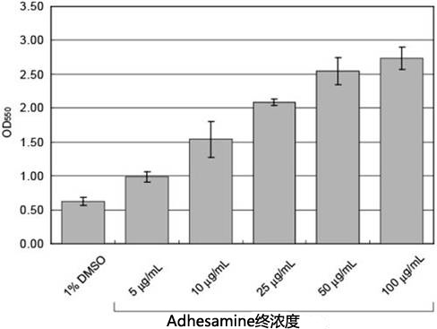
本产品是促进培养容器中的细胞粘附和细胞增殖的低分子化合物。参与细胞表面的硫酸乙酰肝素的结合,显示与纤连蛋白相似的细胞粘附作用。它可以通过添加在培养基中或培养容器的涂层,能使培养容器中的浮游细胞贴壁。附着于含本产品培养容器的细胞,换成不包含本产品的培养基中,即可从培养容器中剥离。HeLa, HEK293, CHO, 小鼠ES细胞等已确认效果1)。此外,培养困难的细胞通过加入本产品的进行培养,细胞增殖有所改善。2)


◆特点


Adhesamine


  ● 具有纤连蛋白相似的细胞粘附作用。

  ● 合成产品感染的危险性小,批次间差异小。

  ● 促进细胞粘附与细胞增殖。

  ● 加入涂料,培养基均可使用。

  ● CAS No.: 462605-73-9

  ● C24H32Cl4N8O2S2 = 670.51


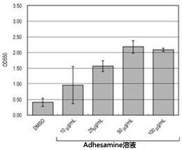
使用例子涂料


Adhesamine


  1. 1mL DMSO 溶解 10 mg Adhesamine(超声波处理分钟)。

  2. 1.配制的作为原液,使用 PBS(-)分别稀释出 10, 25, 50, 100 ug/mL

  3. 各浓度 500 uL Adhesamine 分别注入至24孔培养板,在37℃静置小时。

  4. 抽吸 Adhesamine 溶液,用 PBS(-)清洗。

  5. 各孔播种 0.5×10⁶cells 的 Jurkat 细胞(500ul)。

  6. 37, 5% CO2培养小时⇒ 下图

  7. PBS(-)清洗。

  8. 4%多聚甲醛固定细胞。

  9. 5mg/mL结晶紫染色。

  10. 清洗剩余结晶紫后,干燥。

  11. 用2% SDS溶液洗脱结晶紫,测定 OD550 吸光值。⇒ 右图


      Adhesamine


      使用例子:添加入培养基


      Adhesamine


       1. 1mL DMSO 溶解 10mg Adhesamine分钟左右超声波处理)。

       2. 1.配制的作为原液,使用PBS(-)分别稀释出 0.5, 1.0, 2.5, 5.0 mg/mL

       3. 各浓度 Adhesamine 溶液5uL分别注入至24孔培养板。

       4. 各孔播种 0.5×10cells 的 Jurkat 细胞(500 uL)。

       5. 37, 5% CO2培养小时。⇒ 下图

       6. PBS(-)清洗。

       7. 4%多聚甲醛固定细胞

       8. 5mg/mL结晶紫染色。

       9. 清洗剩余结晶紫后,干燥。

       10. 2% SDS溶液洗脱结晶紫,测定OD550吸光值。右图


      Adhesamine


      使用注意事项

      DMSO 溶解后,溶液有可能析出结晶。析出后不能溶解,建议现配现用。


      ◆参考文献

        1) Yamazoe, S., et al.:, Chem. Biol., 16, 773 (2009).
        2) Hoshino, M., et al.:, Biochem. J., 427, 297 (2010).


      产品编号 产品名称 产品规格 产品等级
      010-23201 Adhesamine 1mg 细胞培养用

      LDH-细胞毒性检测Wako LDH-Cytotoxic Test Wako

      • 产品特性
      • 相关资料
      • Q&A
      • 参考文献

      LDH-细胞毒性检测Wako                              LDH-Cytotoxic Test WakoLDH-细胞毒性检测Wako

      LDH-Cytotoxic Test Wako


      背景


        在日本每年在动物实验中死亡的小鼠和大鼠达850万只。其中以兔子眼睛作为药品和化妆品检测的样本,利用德莱兹法观察角膜和结膜的变化检测毒性·刺激性的实验,是非常残忍的。

        因此,开发了在in vitro条件下的动物实验代替传统动物实验。此方法可检测培养细胞的毒性和细胞增值能力,不仅可测定因试剂破坏细胞而流出的酶量,还可以使色素渗入到活细胞中。比实验动物法实惠且节省操作,可客观地测定其毒性。

        本产品是使用培养细胞对各种试剂的毒性进行简便测定的试剂盒。

        可以直接测定细胞膜受到试剂影响而死亡的细胞中的游离LDH(乳酸脱氢酶),灵敏性高。


      ◆优点


      ● 可用于吸附细胞·浮游细胞

      ● 使用酶测定法进行测定,可以对细胞毒性进行精确的定量测定

      ● 通过调节样品处理时间和显色反应时间可以调节灵敏度

      ● 使用吸光全自动定量绘图酶标仪可以对多数样品进行测定

      ● 测定波长:吸光560 nm


      测定原理


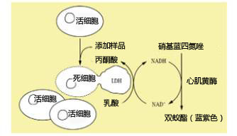
      LDH-细胞毒性检测Wako                              LDH-Cytotoxic Test Wako

        在LDH的作用下乳酸会被丙酮酸氧化,而辅酶会被NADH还原。样品中根据LDH活性按比例生成的NADH会在心肌黄酶的作用下会将硝基蓝四氮唑还原,生成蓝紫色的双蚁酯。在560 nm(±10 nm)下测定这个显色液的吸光度。


      LDH法

      MTT法

      测定对象

      死细胞(&活细胞)

      活细胞

      测定时间(处理样品后)

      1小时内

      4~5小时

      微量毒性的测定

      适用

      适用

      微量死细胞测定时的误差

      结果的客观性


      ◆试剂盒构成


      显色试剂 硝基蓝四氮唑还原

      心肌黄酶,NAD(3.7mg/vial)——————————————5 mL用×10支

      缓冲液 DL-乳酸锂(50mg/mL)——————————————————55 mL×11支

      反应停止液 盐酸(1mol/L)————————————————————55 mL×11支

      96孔微量滴定板—————————————————————————10块(未灭菌)

      ◆使用方法


      实验步骤

      96孔板(灭菌)

      ↓→添加细胞。37℃下过夜

      ↓→PBS清洗

      ↓→添加检测物。37℃,15分钟

      ↓→离心分离

      上清液


      ◆用途


        通过LDH法检测各种试剂毒性的试剂盒。

      参考文献

      [1] Korzeniewski, C. andCallewaert, D. M. : J. Immunol. Methods, 64, 313 (1983)

      [2] Decker, T. andLohmann-Matthes, M. -L. : J. Immunol. Methods, 115, 61 (1988)

      产品编号 产品名称 产品规格 产品等级
      299-50601 LDH-Cytotoxic Test Wako
      LDH细胞毒性检测试剂盒
      960次用 细胞毒性测定用

      台盼蓝溶液

      • 产品特性
      • 相关资料
      • Q&A
      • 参考文献

      台盼蓝溶液台盼蓝溶液


        台盼蓝是用于判断活细胞的细胞染色试剂。死亡细胞会被染成绿色,活细胞不会被染色。因此,可根据染色后细胞的颜色判断其生死,也可以利用血球计数板计算细胞的数量。
        本产品由 D-PBS(-) 调制而成。

      〔细胞种类〕 人类急性单核性白血病细胞系
      〔混合比率〕 细胞悬浮液:台盼蓝溶液 = 1:1


      相关产品


      ◆StenSure®系列


        以下产品使用小鼠ES细胞D3系进行质量测试。

        ● StemSure®D-MEM・巯基乙醇・硫代甘油

        ● StemSure®明胶溶液

        ● StemSure®血清替代品

        ● StemSure®冷冻保存溶液

        ● StemSure®LIF、小鼠,、重组体、溶液


      产品编号

      产品名称

      规格

      容量

      197-17275

      StemSure®  D-MEM with Phenol Red and Sodium Pyruvate

      用于培养细胞

      500 mL

      197-16775

      StemSure®   Serum Replacement

      用于培养细胞

      500 mL

      198-15781

      StemSure® 10mmol/l 2-Mercaptoethanol Solution(×100)

      用于培养细胞

      100 mL

      195-15791

      StemSure® 50mmol/l Monothioglycerol Solution(×100)
       可代替2ME使用的还原剂。不属于有毒物。

      用于培养细胞

      100 mL

      190-15805

      StemSure®  0.1w/v% Gelatin Solution

      用于培养细胞

      500 mL

      195-16031

      StemSure® Freezing Medium

      用于培养细胞

      100 mL

      199-16051

      StemSure®  LIF, Mouse, recombinant, Solution

      用于培养细胞

      106 units

       
      ◆ES・iPS 细胞研究试剂
       
        2007年公布iPS细胞建立后,出现了大量关于iPS细胞的文献。在众多文献中,列出了有关ES细胞・iPS细胞的未分化能维持及分化诱导的低分子化学物。<链接>
       
       
      神经干细胞补充产品
       

        本产品是用于培养神经干细胞的血清替代品。以下列出的两种产品分别为转铁蛋白(脱铁)产品及转铁蛋白(全铁)。一般产品都会含有转铁蛋白(全铁),而添加了含有转铁蛋白(脱铁)的N2补充产品,会有利于某些细胞种类的干细胞繁殖。

        N2补充产品[含有转铁蛋白(脱铁)]

        N2补充产品[含有转铁蛋白(全铁)]


      产品编号

      产品名称

      规格

      容量

      141-09041

      N2 Supplement with Transferrin(Apo)(×100)

      用于培养细胞

      5 mL

      141-08941

      N2 Supplement with Transferrin(Holo)(×100)

      用于培养细胞

      5 mL


      产品编号 产品名称 产品规格 产品等级
      207-17081 0.4w/v% Trypan Blue Solution 100 mL 用于细胞染色

      Stellar化学转化感受态细胞酶试剂盒Takara

      上海金畔生物科技有限公司代理Takara酶试剂盒全线产品,欢迎访问官网了解更多产品信息。

      Stellar化学转化感受态细胞
      品牌 Code No. 产品名称 包装量 价格(元) 说明书 数量
      Clontech 636763 Stellar Competent Cells 10 x 100 μl ¥2,315 Stellar化学转化感受态细胞 Stellar化学转化感受态细胞 Stellar化学转化感受态细胞
      Clontech 636766 Stellar Competent Cells 50 x 100 μl ¥8,189 Stellar化学转化感受态细胞 Stellar化学转化感受态细胞 Stellar化学转化感受态细胞
      Clontech 636767 Stellar Competent Cells (96-well plate) 96 x 20 μl ¥5,518 Stellar化学转化感受态细胞 Stellar化学转化感受态细胞 Stellar化学转化感受态细胞
      Clontech 636764 Stellar Competent Cells (dam-/dcm-) 10 transformations ¥2,532 Stellar化学转化感受态细胞 Stellar化学转化感受态细胞 Stellar化学转化感受态细胞
      收藏产品 加入购物车

      无标题文档


      Stellar化学转化感受态细胞 Stellar化学转化感受态细胞 Stellar化学转化感受态细胞 Stellar化学转化感受态细胞 Stellar化学转化感受态细胞
       
      Stellar化学转化感受态细胞
      Stellar化学转化感受态细胞  
      Stellar化学转化感受态细胞
       
       
      Stellar Competent Cells属于E. coli HST08细胞系,具有很高的转化效率,可用于多种应用:cDNA或基因组DNA文库的制备,长片段基因组文库的构建,亚克隆,甚至甲基化DNA的克隆。Stellar Competent Cells缺失切断外源甲基化DNA的基因簇(mrr-hsdRMS-mcrBC and mcrA),所以可用于克隆甲基化DNA。 转化包含lacZ alpha基因的载体时,也可以使用Stellar Competent Cells进行蓝白筛选,例如alpha互补。 Stellar Competent Cells是Clontech's In-Fusion Cloning Kits配套使用的理想选择。
       
      Stellar Competent Cells (dam/dcm)
      Stellar Competent Cells (dam/dcm)属于E. coli HST04细胞系,缺失了甲基化DNA所必须的遗传因子(damdcm)。通常被dam或dcm甲基化而抑制的限制酶可以切断由该产品制备的质粒。
       
      产品详情请点击:Stellar化学转化感受态细胞


      页面更新:2019-11-05 09:50:57

      PrimeSurface 低吸附细胞培养板 PrimeSurface 35mm dish

      • 产品特性
      • 相关资料
      • Q&A
      • 参考文献

      PrimeSurface 低吸附细胞培养板                              PrimeSurface 35mm dish低吸附相关试剂

      PrimeSurface 低吸附细胞培养板


      PrimeSurface® 是采用 sumiron 公司的低吸附蛋白质处理产品(Proteosave® SS)的技术制成的低吸附细胞处理产品。使用该容器培养细胞更容易形成细胞球集落。

      ◆特长


      ● 能够简单地形成细胞集落

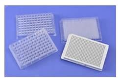
            将细胞播种在96、384多孔板中,静置培养就能简单地得到细胞集落。 

      ● 细胞集落大小均一 

            抑制细胞吸附在培养面,集落的形成率得到了提高,能够在细胞形态平整、均一的状态下培养。

      ● 适用于细胞分化的研究 

            能够将ES细胞变成胚叶体(EB体)后直接添加分化诱导试剂 。

      ● 适合用于使用细胞球进行抗癌剂的筛选 

            进行3D培养,与常规的单层培养相比培养环境更加接近生物体。


      PrimeSurface 低吸附细胞培养板                              PrimeSurface 35mm dish


      ◆用途


      用于再生医疗和药物研发中


      ◆细胞培养


      ●    ES细胞·IPS细胞·骨髓间充质干细胞的分化诱导

      ●    使用细胞的3D模型进行药物研发筛选


      ◆产品概要

      产品编号

      产品名称

      规格

      培养面积

      容量

      包装

      MS-9035XZ

      PrimeSurface® 培养皿35mm

      外寸35Φ×14(H)mm

      9 cm2

      5包·50/case

      MS-9060XZ

      PrimeSurface® 培养皿60mm

      外寸60Φ×15(H)mm

      21 cm2

      10包·100/case

      MS-9090XZ

      PrimeSurface® 培养皿90mm

      外寸90Φ×20(H)mm

      57 cm2

      10包·50/case

      MS-9024XZ

       Sumiron Celltight 板24F

      24孔·平底

      1.8 cm2 

      3.4 mL/well

      1包·10/case


      注:已进行放射线灭菌  保存温度:室温 有效期:制造后两年


      产品编号

      产品名称

      孔数

      孔底形状

      容量

      包装

      MS-9384UZ

      PrimeSurface® 384U多孔板

      384

      U底

      0.1 mL

      1/包・20/case

      MS-9384WZ

      PrimeSurface® 384U白色多孔板

      384

      U底

      0.1 mL

      1/包・20/case

      MS-9096VZ

      PrimeSurface® 96V多孔板

      96

      V底

      0.3 mL

      1/包・20/case

      MS-9096MZ

      PrimeSurface® 96M多孔板

      96

      纺锤底

      0.2 mL

      1/包・20/case

      MS-9096UZ

      PrimeSurface® 96U多孔板

      96

      U底

      0.3 mL

      1/包・20/case

      MS-9096WZ

      PrimeSurface® 96U白色多孔板

      96

      U底

      0.3 mL

      1/包・20/case


      注:已进行放射线灭菌  保存温度:室温  有效期:制造后两年


      <span style="font-family: 微软雅黑, "Microsoft YaHei"; font-size: 14px;"><span style="font-size: 14px; font-family: 微软雅黑, "Microsoft YaHei";"><span style="font-family: 微软雅黑, 'Microsoft YaHei'; font-size: 14px;">无标题文档</span></span></span>

      ◆实验例子


      在用球体制作工程构建三维结构体时使用了 Cyfuse 的生物3D打印机 Regenova®

      Regenova® 是先进的机器人系统之一,它通过将球体固定在剑山,再按照3D设计制作出三维结构体。下面为大家介绍使用了该系统的神经三维结构体和使用了间充质干细胞的三维结构体的实验例子。


      神经三维结构体


      使用细胞:人iPSC来源神经前驱细胞

      播种数:4×10⁴ cells/well

      培养基:神经细胞用培养基

      培养板培养天数:2天

      制作出的三维结构体的形状、尺寸:3×3×2

      用于三维结构体的细胞块个数:18个

      积层后的培养天数:9天后拔出剑山


      PrimeSurface 低吸附细胞培养板                              PrimeSurface 35mm dish

      PrimeSurface 低吸附细胞培养板                              PrimeSurface 35mm dish

      PrimeSurface 低吸附细胞培养板                              PrimeSurface 35mm dish

      细胞块

      3D打印后

      拔出剑山后的三维结构体

      使用间充质干细胞的三维结构体


      使用细胞:人脂肪组织来源间充质干细胞

      播种数:5×10³ cells/well

      培养基:间充质干细胞用培养基

      培养板培养天数:2天

      制作出的三维结构体的形状、尺寸:用48个细胞块组成环状×10层

      用于三维结构体的细胞块个数:480个

      积层后的培养天数:6天后拔出剑山


      PrimeSurface 低吸附细胞培养板                              PrimeSurface 35mm dish

      PrimeSurface 低吸附细胞培养板                              PrimeSurface 35mm dish

      PrimeSurface 低吸附细胞培养板                              PrimeSurface 35mm dish

      PrimeSurface 低吸附细胞培养板                              PrimeSurface 35mm dish

      细胞块

      3D打印后(上面)

      3D打印后(侧面)

      拔出剑山后


      使用 PrimeSurface® 96U 多孔板的培养例子


      小鼠ES细胞培养


      播种数:750 cells/well
      培养基:DMEM+4.5 mg/mL Glc.
      添加物:15%灭活 FCS
                    2 mM L-谷氨酰胺
                    1%非必须氨基酸
                    110 μM 2-巯基乙醇  
      培养日数:3日

      PrimeSurface 低吸附细胞培养板                              PrimeSurface 35mm dish

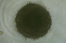
      没有细胞吸附在 PrimeSurface® 96U 多孔板上,也没有凌乱的细胞集落,已经形成均一。

      PrimeSurface 低吸附细胞培养板                              PrimeSurface 35mm dish

      一般市面上贩卖的漂浮培养用多孔板,细胞紧密粘附或者死亡。

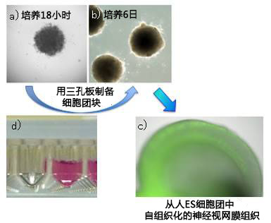
      使用PrimeSurface® 96U多孔板的细胞分化诱导实验例子


      使用PrimeSurface® MS-9096V将人ES细胞集落导入自身组织化神经网膜组织


      培养器:PrimeSurface® MS-9096V

      使用细胞:人ES(KhES-1株)

      播种密度:9000 cells/well

      使用培养基:GMEM+KSR+NEAA+2ME+ 20uM Y-27632

      培养条件:5% CO2,37℃


      PrimeSurface 低吸附细胞培养板                              PrimeSurface 35mm dish


      【数据提供】

      ・照片a)~c) 

       理化学研究所 发生与再生科学综合研究中心 干细胞研究支援开发室


      【参考文献】

      ・Self-Formation of Optic Cups and Storable Stratified Neural Retina from Human ESCs

       NakanoT, Ando S, Takata N, Kawada M, Muguruma K, Sekiguchi K, Saito K, Yonemura S, Eiraku M, Sasai Y

       Cell Stem Cell, 10 (6), 771-785 (2012)


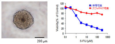
      使用PrimeSurface® 进行抗癌药药效测试实验


      细胞:MCF-7(人乳腺癌细胞)

      试剂:5-Fluorouracil(5-FU)


      PrimeSurface 低吸附细胞培养板                              PrimeSurface 35mm dish


      【数据提供】

      ・近畿大学医学部 基因组生物学教室 西尾研究室


      在小鼠 ES 细胞形成球体引起心肌细胞分化的过程中使用 DS Pharma Biomedical 的心肌分化毒性(发生毒性)评价试剂盒。


      POCA® Hand1-EST


      POCA® Hand1-EST 是一种使用 PrimeSurface,在待测物存在下的培养中将检测小鼠ES细胞的心肌分化是否正常作为标记基因活性的指标的方法。


      使用细胞:小鼠ES细胞(Hand1-ES细胞)

      播种数:750 cells/well

      培养基:心肌分化培养基

      培养板培养天数:5天


      PrimeSurface 低吸附细胞培养板                              PrimeSurface 35mm dish

      研究领域

      使用型号

      参考文献

      视网膜

      研究

      MS-9096V

      KUWAHARA, Atsushi, et al. Generation of a ciliary margin-like stem cell niche from self-organizing human retinal tissue. Nature communications, 2015, 6.: 1-15

      MS-9096V

      TANAKA, T., et al. Generation of retinal ganglion cells with functional axons from human induced pluripotent stem cells. Sci Rep, 2015, 5. 8344.

      MS-9096U

      EIRAKU, Mototsugu and SASAI, Yoshiki Mouse embryonic stem cell culture for generation of three-dimensional retinal and cortical tissues. Nature protocols,

      2012, 7. 1: 69-79. 

      MS-9096V

      NAKANO, Tokushige, et al. Self-Formation of Optic Cups and Storable Stratified Neural Retina from Human ESCs. Cell Stem Cell, 2012, 10. 6: 771-785.

       MS-9096V

      GAO, Lixiong, et al. Intermittent high oxygen influences the formation of neural retinal tissue from human embryonic stem cells.Scientific Reports, 2016, 6.

      神经科学

      研究

      MS-9096V

      MUGURUMA, Keiko, et al. Self-Organization of Polarized Cerebellar Tissue in 3D Culture of Human Pluripotent Stem Cells. Cell Reports, 2015, 10:537-550

      MS-9096V

      BAMBA, Y., et al. Differentiation, polarization, and migration of human induced pluripotent stem cell-derived neural progenitor cells co-cultured with a human glial cell   line with radial glial-like characteristics. Biochem Biophys Res Commun,   2014, 447. 4: 683-688. 

      MS-9096 U, M or V

      MINAMINO, Yuki, et al. Isolation and Propagation of Neural Crest Stem Cells from Mouse Embryonic Stem Cells via Cranial Neurospheres. Stem cells and development, 2014, 24.2:   172-181

      MS-9096V

      KADOSHIMA, T., et al. Self-organization of axial polarity, inside-out layer pattern, and species-specific progenitor dynamics in human ES cell-derived neocortex. Proceedings of the National Academy of Sciences of the United States of   America, 2013, 110. 50: 20284-20289. 

      MS-9096 U, M or V

      OGAWA, Yasuhiro, et al. Impaired neural differentiation of induced pluripotent stem cells generated from a mouse model of Sandhoff disease. PLoS ONE, 2013, 8. 1: e55856.

      MS-9035X

      GOMI, Masanori, et al. Functional recovery of the murine brain ischemia model using human induced pluripotent stem cell-derived telencephalic progenitors. Brain research, 2012, 1459. 52-60. 

      MS-9096 U, M or V

      NASU, Makoto, et al. Robust formation and maintenance of continuous stratified cortical   neuroepithelium by laminin-containing matrix in mouse ES cell culture. PLoS ONE, 2012, 7. 12: e53024.

      MS-9096 U

      DANJO, T., et al. Subregional specification of embryonic stem cell-derived ventral telencephalic tissues by timed and combinatory treatment with extrinsic signals. The Journal of neuroscience : the official journal of the Society   for Neuroscience, 2011, 31. 5: 1919-1933.

      MS-9096 U

      KANEMURA, Yonehiro Development of cell-processing systems for human stem cells (neural stem cells, mesenchymal stem cells, and iPS cells) for regenerative medicine. The   Keio journal of medicine, 2010, 59. 2: 35-45.

      MS-9096 U, M or V

      MS-9035X, MS-9060X

      or

      MS-9090X

      FUKUSUMI, Hayato, et al. (2016). Establishment of human neural progenitor cells from human induced pluripotent stem cells with diverse tissue. Stem cells international: 1-10.

       MS-9096 U, M or V

      RAASCH, Martin, et al. (2016). An integrative microfluidically supported in vitro model of an endothelial barrier combined with cortical spheroids simulates effects of neuroinflammation in neocortex development. Biomicrofluidics. 10: 044102.

      MS-9096 M

      ISODA, Miho, et al. (2016). Robust production of human neural cells by establishing neuroepithelial-like stem cells from peripheral blood mononuclear cell-derived feeder-free iPSCs under xeno-free conditions. Neuroscienc   Research.

      MS-9035X, MS-9060X

      or

      MS-9090X

      BAMBA, Yohei, et al. (2016). In vitro characterization of neurite extension using induced pluripotent stem cells derived from lissencephaly patients with TUBA1A missense mutations. Molecular brain.

      MS-9096V

      SAKAGUCHI, Hideya, et al. (2015). Generation of functional hippocampal neurons from self-organizing human embryonic stem cell-derived dorsomedial telencephalic tissue. Nature communications. 6: 1-11.

      MS-9096V

      MUGURUMA, Keiko, et al. Self-Organization of Polarized Cerebellar Tissue in 3D Culture of Human Pluripotent Stem Cells. Cell Reports, 2015, 10:537-550

      KAMIYA, Daisuke, et al. Intrinsic transition of embryonic stem-cell differentiation into neural progenitors. Nature, 2011, 470. 7335: 503-509.

      心肌细胞

      研究和心脏研究

      MS-9096V

      TAKASHIMA, Yasuhiro, et al. Resetting transcription factor control circuitry toward ground-state pluripotency in human. Cell, 2014, 158. 6: 1254-1269.

      MS-9035X

      OTSUJI, Tomomi G, et al. Dynamic link between histone H3 acetylation and an increase in the functional characteristics of human ESC/iPSC-derived cardiomyocytes. PLoS ONE, 2012, 7. 9: e45010.

      MS-9096U

      SATOSHI, Yasuda, et al. AW551984: a novel regulator of cardiomyogenesis in pluripotent embryonic cells. Biochemical Journal, 2011, 437. 2: 345-355.

      MS-9096U

      YASUDA, S., et al. A novel regulator of cardiomyogenesis in pluripotent embryonic cells. The Biochemical journal, 2011, 437. 2: 345-355.

      MS-9096U

      OTSUJI, Tomomi G, et al. Progressive maturation in contracting cardiomyocytes derived from human embryonic stem cells: Qualitative effects on electrophysiological responses to drugs. Stem cell research, 2010, 4. 3: 201-213.

      MS-9035X, MS-9060X or MS-9090X

      YAMAUCHI, Kaori, et al. Cardiomyocytes develop from anterior primitive streak cells induced by β‐catenin activation and the blockage of BMP signaling in hESCs. Genes to Cells, 2010, 15. 12: 1216-1227.

      MS-9096V

      GUO, Ge, et al. (2016). Naive pluripotent stem cells derived directly from isolated cells of the human innercell mass. Stem Cell Reports. 6: 437-446.

      MS-9096 U

       MS-9035X, MS-9060X

      or

      MS-9090X

      NOGUCHI, Ryo, et al. (2016). Development of a three-dimensional pre-vascularized scaffold-freecontractile cardiac patch for treating heart disease. The Journal of Heart and Lung Transplantation. 35: 137-145.

       MS-9096

      NOGUCHI, Ryo, et al. Development of a Three-Dimensional Prevascularized Scaffold-Free Contractile Cardiac Patch for Treating Heart Disease. The Journal of Heart and Lung Transplantation, 2015,

      肝细胞

      研究

      MS-9096 U, M or V

      ISHII, Takamichi (2012).   Differentiation of Human Embryonic Stem Cells into Functional Hepatocyte-Like Cells (Method). Stem Cells and Cancer Stem Cells, Volume 2, Springer:   43-49. 

      MS-9096 U, M or V

      ISHII, Takamichi et al. (2012). Hepatic Maturation of hES Cells by Using a Murine Mesenchymal Cell Line Derived from Fetal Livers.Human Embryonic and Induced Pluripotent Stem Cells, Springer: 397-403.

      MS-9096U

      ISHII, Takamichi, et al. In vitro hepatic maturation of human embryonic stem cells by using a mesenchymal cell line derived from murine fetal livers. Cell and tissue research, 2010, 339. 3: 505-512. 

      MS-9096U

      YANAGIDA, Ayaka, et al. Liver maturation deficiency in p57 Kip2-/-mice occurs in a hepatocytic p57 Kip2 expression-independent manner. Developmental biology, 2015,

      牙科

      研究

      MS-9035X, MS-9060X or MS-9090X

      OZEKI, Nobuaki, et al. Differentiation of Human Skeletal Muscle Stem Cells into Odontoblasts Is Dependent on Induction of α1 Integrin Expression.Journal of Biological Chemistry, 2014, 289. 20: 14380-14391.

      MS-9096 U, M or V

      YAMAMOTO, Mioko, et al. Three-dimensional spheroid culture promotes odonto/osteoblastic differentiation of dental pulp cells.Archives of oral biology, 2014, 59. 3: 310-317.

      生精小管

      研究

      MS-9096V

      YOKONISHI, T., et al. In Vitro Reconstruction of Mouse Seminiferous Tubules Supporting Germ Cell Differentiation. Biol Reprod, 2013, 89. (1):15: 1–6.

      iPS细胞

      生成

      MS-9035X

      OHNISHI, Hiroe, et al. A comparative study of induced pluripotent stem cells generated from frozen, stocked bone marrow‐and adipose tissue‐derived mesenchymal stem cells. Journal of tissue engineering and regenerative medicine, 2012, 6. 4: 261-271. 1. 

      MS-9035X

      AOKI, T., et al. Generation of induced pluripotent stem cells from human adipose-derived stem cells without c-MYC. Tissue engineering. Part A, 2010, 16. 7: 2197-2206.

      MS-9035X, MS-9060X or MS-9090X

      ODA, Y., et al. Induction of pluripotent stem cells from human third molar mesenchymal stromal cells. J Biol Chem, 2010, 285. 38: 29270-29278.

      OHNISHI, Hiroe, et al. Generation of Xeroderma Pigmentosum-A Patient-Derived Induced Pluripotent Stem Cell Line for Use As Future Disease Model. Cellular Reprogramming (Formerly" Cloning and Stem Cells"), 2015, 17. 4: 268-274.

      EST(胚胎干细胞试验)

      MS-9096W

      SUZUKI, N., et al. Evaluation of novel high-throughput embryonic stem cell tests with new molecular markers for screening embryotoxic chemicals in vitro. Toxicological sciences : an official journal of the Society of Toxicology, 2011, 124. 2: 460-471.

      MS-9096U

      NAGAHORI, H., et al. (2016). Prediction of in vivo developmental toxicity by combination of Hand1-Luc embryonic stem cell test and metabolic stability test with clarification of metabolically inapplicable candidates. Toxicol Lett. 259: 44-51.

       MS-9096U

      YU, Ruoxing, et al. (2015). A Modified Murine Embryonic Stem Cell Test for Evaluating the Teratogenic Effects of Drugs on Early Embryogenesis. PLoS ONE. 10: e0145286.

      MS-9096W

      COZ, Florian Le, et al. (2015). Hand1-Luc Embryonic Stem Cell Test (Hand1-Luc EST): A novel rapid and highly reproducible in vitro test for embryotoxicity by measuring cytotoxicity and differentiation toxicity using engineered mouse ES cells. The Journal of Toxicological Sciences. 40: 251-261.

      骨和软骨研究

      MS-9096 U

      HINO, Kyosuke, et al. (2015). Neofunction of ACVR1 in fibrodysplasia ossificans progressiva. Proceedings ofthe National Academy of Sciences. 112: 15438-15443.

      MS-9096 U, M or V

      MURATA, Daiki, et al. A preliminary study of osteochondral regeneration using a  scaffold-free threedimensional construct of porcine adipose tissue-derived mesenchymal stem cells. Journal of orthopaedic surgery and research, 2015, 10. 1: 1-12.

       MS-9096 U, M or V

      FUJIMOTO, Mai, et al. Establishment of a novel model of chondrogenesis using murine embryonic stem cells carrying fibrodysplasia ossificans progressiva-associated mutant ALK2. Biochemical and Biophysical Research Communications,

      2014, 455. 3: 347-352. 1.

      MS-9096 U

      ISHIHARA, Kohei, et al. Simultaneous regeneration of full-thickness cartilage and subchondral bone defects in vivo using a three-dimensional scaffold-free autologous construct derived from high-density bone marrow-derived mesenchymal stem cells. J Orthop Surg Res, 2014, 9. 1: 98. 1.

      血管

      研究

       MS-9096 U

      MS-9035X, MS-9060X

      or

      MS-9090X

      KAGEYAMA, Tatsuto, et al. (2016). In situ crosslinkable gelatin-CMC hydrogels designed for rapid engineering of perfusable vasculatures. ACS Biomaterials Science & Engineering.

      MS-9096U

      ITOH, M., et al. Scaffold-Free Tubular Tissues Created by a Bio-3D Printer Undergo Remodeling and Endothelialization when Implanted in Rat Aortae.PLoS ONE,

      2015, 10. 9: e0136681.

      胰岛细胞移植

      MS-9096 U

      NAKAMURA, Kentaro, et al. (2016). Introduction to a new cell transplantation platform via recombinant peptide petaloid pieces and its application to islet transplantation with mesenchymal stem cells. Transplant International. 29: 1039-1050.

      骨髓

      研究

       MS-9096 U

      SAYO, Kanae, et al. (2016). Fabrication of bone marrow-like tissue in vitro from dispersed-state bone marrow cells. Regenerative Therapy. 3: 32-37.

      其它

      MS-9035X, MS-9060X

      or

      MS-9090X

      ITO, Yoshitaka, et al. (2015). Establishment of Tsc2deficient rat embryonic stem cells. International journal of oncology. 46: 1944-1952.

       MS-9096 U, M or V

      OGAWA, Yasuhiro, et al. (2015). Induced Pluripotent Stem Cells Generated from P0-Cre; Z/EG Transgenic Mice. PLoS ONE. 10: e0138620.

      MS-9096U

      IMAI, Hiroyuki, et al. Tetraploid Embryonic Stem Cells Maintain Pluripotency and Differentiation Potency into Three Germ Layers. PLoS ONE, 2015, 10. 6: e0130585.

      MS-9096U, MS-9096M

      or

      MS-9096V

      MITSUI, Kaoru, et al. Conditionally replicating adenovirus prevents pluripotent stem cell–derived teratoma by specifically eliminating undifferentiated cells. Molecular Therapy. Methods & Clinical Development, 2015, 2. 15026.

      MS-9096U

      ZHOU, Yuanshu, et al. Metabolic suppression during mesodermal differentiation of embryonic stem cells identified by single-cell comprehensive gene expression analysis. Molecular BioSystems, 2015, 11. 9: 2560-2567.

      PrimSurface® 肿瘤研究参考文献


        1. 

      ROTEM, Asaf, et al. Alternative to the soft-agar assay that permits high-throughput   drug and genetic screens for cellular transformation. Proceedings of the National Academy of Sciences, 2015, 1-6

        2.

      OHNISHI, Ken, et al. Plastic induction of CD133AC133-positive cells in the microenvironment of glioblastoma spheroids.International Journal of Oncology, 2014, 45.2: 581-586.

        3.

      KODAMA, Tatsushi, et al. A Novel Mechanism of EML4-ALK Rearrangement Mediated by Chromothripsis in a Patient-Derived Cell Line. Journal of Thoracic Oncology, 2014, 9.11: 1638-1646.

        4.

      SHIMOZATO, O., et al. Receptor-type protein tyrosine phosphatase κ directly dephosphorylates CD133 and regulates downstream AKT activation. Oncogene, 2014.

        5.

      MIKHAIL, Andrew S., et al. Image-based analysis of the size-and time-dependent penetration of polymeric micelles in multicellular tumor spheroids and tumor xenografts. International journal of pharmaceutics, 2014, 464.1: 168-177.

        6.

      MORI, Masamichi, et al. The Selective Anaplastic Lymphoma Receptor Tyrosine Kinase Inhibitor ASP3026 Induces Tumor Regression and Prolongs Survival in Non–Small Cell Lung Cancer Model Mice. Molecular cancer therapeutics, 2014, 13.2: 329-340.

        7.

      AKIMOTO, Miho, et al. An inhibitor of HIF-α subunit expression suppresses hypoxia-induced dedifferentiation of human NSCLC into cancer stem cell-like cells. World, 2013, 3.4: 41-45.

        8.

      GOUDARZI, Houman, et al. Hypoxia affects in vitro growth of newly established cell lines from patients with malignant pleural mesothelioma. Biomedical Research, 2013, 34.1:   13-21.

        9.

      KATO, Takuma; TANAKA, Masakazu; OBA, Makoto. Protein Transfection Study Using Multicellular Tumor Spheroids of Human Hepatoma Huh-7 Cells. PloS one, 2013, 8.12: e82876.

      10.

      MIKHAIL, Andrew S.; EETEZADI, Sina; ALLEN, Christine. Multicellular tumor spheroids for evaluation of cytotoxicity and tumor growth inhibitory effects of nanomedicines in vitro: A comparison of docetaxel-loaded block copolymer   micelles and Taxotere®.PloS one, 2013, 8.4: e62630.

      11.

      SATO, Shuji, et al. Identification of the cancer cell proliferation and survival functions of proHB-EGF by using an anti-HB-EGF antibody. PloS one, 2013, 8.1: e54509.

      12.

      ISHII, Genichiro, et al. Morphophenotype of floating colonies derived from a single cancer cell has a critical impact on tumor‐forming activity. Pathology international, 2013, 63.1: 29-36.

      13.

      OYANAGI, Jun, et al. Epithelial-mesenchymal transition stimulates human cancer cells   to extend microtubule-based invasive protrusions and suppresses cell growth in collagen gel. PloS one, 2012, 7.12: e53209.

      14.

      UNO,   Makiko, et al. Identification of physiologically active substances as novel ligands for MRGPRD. BioMed Research International, 2012, 2012.

      15.

      KONISHI, Hiroaki, et al. PEGylated liposome IHL-305 markedly improved the survival of ovarian cancer peritoneal metastasis in mouse. BMC cancer, 2012, 12.1: 462.

      16.

      NISHIMURA, Satoko, et al. MRGD, a MAS-related G-protein coupled receptor, promotes tumorigenisis and is highly expressed in lung cancer. PloS one, 2012, 7.6: e38618.

      17.

      SAKAMOTO, Hiroshi, et al. CH5424802, a selective ALK inhibitor capable of blocking the resistant gatekeeper mutant. Cancer cell, 2011, 19.5: 679-690.

      18.

      KUNITA, Akiko, et al. Podoplanin is regulated by AP-1 and promotes platelet aggregation and cell migration in osteosarcoma. The American journal of pathology, 2011, 179.2: 1041-1049.

      19.

      SAKUMA, Yuji, et al. WZ4002, a third-generation EGFR inhibitor, can overcome anoikis resistance in EGFR-mutant lung adenocarcinomas more efficiently than Src inhibitors. Laboratory Investigation, 2011, 92.3: 371-383.

      20.

      SHIMIZU, Yutaka, et al. Dienogest, a synthetic progestin, inhibits prostaglandin E2 production and aromatase expression by human endometrial epithelial cells in a spheroid culture system. Steroids, 2011, 76.1: 60-67.

      21.

      KOSHIKAWA, Naohiko, et al. Proteolytic activation of heparin‐binding EGF‐like growth factor by membrane‐type matrix metalloproteinase‐1 in ovarian carcinoma cells. Cancer science, 2011, 102.1: 111-116.

      22.

      SAKAMOTO, Hiroshi, et al. CH5424802, a selective ALK inhibitor capable of blocking the resistant gatekeeper mutant. Cancer cell, 2011, 19.5: 679-690.

      23.

      YAMAGUCHI,   Shigeru, et al. Novel Photodynamic Therapy Using Water‐dispersed TiO2–Polyethylene Glycol Compound: Evaluation of Antitumor Effect on Glioma Cells and Spheroids In Vitro. Photochemistry and photobiology,   2010, 86.4: 964-971.

      24.

      MATSUYAMA, Masahiro, et al. Reduced CD73 expression and its association with altered purine nucleotide metabolism in colorectal cancer cells robustly causing liver metastases. Oncology letters, 2010, 1.3: 431-436.

      25.

      KOGASHIWA, Yasunao, et al. Docetaxel suppresses invasiveness of head and neck cancer cells in vitro. Cancer science, 2010, 101.6: 1382-1386.

      26.

      HAN, Muri, et al. Enhanced percolation and gene expression in tumor hypoxia by PEGylated polyplex micelles. Molecular Therapy, 2009, 17.8: 1404-1410.

      27.

      WATANABE, Y., et al. A novel translational approach for human malignant pleural mesothelioma: heparanase-assisted dual virotherapy. Oncogene, 2009, 29.8: 1145-1154.

      28.

      KUNITA, Akiko, et al. The platelet aggregation-inducing factor aggrus/podoplanin promotes pulmonary metastasis. The American journal of pathology,   2007, 170.4: 1337-1347.

      29.

      HAN,  Muri, et al. Transfection study using multicellular tumor spheroids for screening non-viral polymeric gene vectors with low cytotoxicity and high transfection efficiencies. Journal of Controlled Release, 2007, 121.1: 38-48.

      30.

      KESSEL, Sarah, et al. (2016). High-Throughput 3D Tumor Spheroid Screening Method for Cancer Drug Discovery Using Celigo Image Cytometry. Journal of laboratory automation: 2211068216652846.

      31.

      OZAWA, Masayuki (2015). The N-cadherin cytoplasmic domain confers anchorage-independent growth and the loss of contact inhibition. Scientific Reports. 5: 1-16.

      32.

      TARIQ, Mohammad, et al. (2016). Eukaryotic translation initiation factor 5A (eIF5A) is essential for HIF-1α activation in hypoxia. Biochemical and Biophysical Research Communications. 470: 417-424.

      33.

      TERASHIMA, Jun, et al. (2016). VEGF expression is regulated by HIF-1α and ARNT in 3D KYSE-70, esophageal cancer cell spheroids. Cell biology international.

      34.

      MASUDA, Mari, et al. (2016). TNIK inhibition abrogates colorectal cancer stemness. Nature communications. 7: 12586.

      35.

      SUDA, Yoshitaka, et al. (2016). Clonal heterogeneity in osteogenic potential of lung cancer-associated fibroblasts: promotional effect of osteogenic progenitor cells on cancer cell migration. Journal of cancer research and clinical oncology: 1-12.

      36.

      EETEZADI, Sina, et al. (2015). Effects of Doxorubicin Delivery Systems and Mild Hyperthermia on

      Tissue Penetration in 3D Cell Culture Models of Ovarian Cancer Residual Disease. Molecular pharmaceutics. 12: 3973-3985.

      37.

      MISU, Masayasu, et al. Effects of Wnt-10b on proliferation and differentiation of murine melanoma cells.Biochemical and Biophysical Research Communications, 2015, 463. 4: 618-623.

      38.

      NAKANISHI, Yoshito, et al. Mechanism of oncogenic signal activation by the novel fusion kinase FGFR3– BAIAP2L1.Molecular cancer therapeutics, 2015, 14. 3: 704-712.

      39.

      NOZAWA-SUZUKI, Noriko, et al. The inhibitory effect of hypoxic cytotoxin on the expansion of cancer stem cells in ovarian cancer.Biochemical and Biophysical Research Communications, 2015, 457. 4: 706-711.

      40.

      YONESAKA, Kimio, et al. (2015). The pan-HER family tyrosine kinase inhibitor afatinib overcomes HER3 ligand heregulin-mediated resistance to EGFR inhibitors in non-small cell lung cancer. Oncotarget.    6: 33602.

      41.

      YONESAKA, K, et al. Anti-HER3 monoclonal antibody patritumab sensitizes refractory non-small cell lung cancer to the epidermal growth factor receptor inhibitor erlotinib.Oncogene, 2015

      42.

      YOSHIDA, Ryohei, et al. EGFR tyrosine kinase inhibitors combined with cytotoxic drugs for treatment of NSCLC with EGFR gene mutations: Efficacy and mechanisms.Cancer research, 2015, 75. 15 Supplement: 3501-3501.

      43.

      KODAMA, Tatsushi, et al. Alectinib shows potent antitumor activity against RET-rearranged non–small cell lung cancer.Molecular cancer therapeutics, 2014, 13. 12: 2910-2918.

      44.

      寺島潤 (2014). 肝がん 3D 細胞塊における薬物代謝酵素の不均一な発現. バイオテック 2014.

      45.

      中野洋文 難治性癌・癌肝細胞の三次元培養薬剤スクリーニング.Bio Garage, 2014, 22. 13.

      46.

      BRESLIN, Susan and O’DRISCOLL, Lorraine Three-dimensional cell culture: the missing link in drug discovery.Drug Discovery Today, 2013, 18. 5: 240-249.

      47.

      ISHII, Genichiro, et al. Morphophenotype of floating colonies derived from a single cancer cell has a critical impact on tumor-forming activity.Pathology International, 2013, 63. 1: 29-36.

      48.

      KATO, Takuma, et al. Protein Transfection Study Using Multicellular Tumor Spheroids of Human Hepatoma Huh-7 Cells.PLoS ONE, 2013, 8. 12: e82876.

      49.

      KESSEL, Sarah, et al. (2013). Progressing 3D Spheroid Analysis into a HTS Drug Discovery Method. Molecular biology of the cell, AMER SOC CELL BIOLOGY 8120 WOODMONT AVE, STE 750, BETHESDA, MD 20814-2755 USA.

      50.

      MASUDA, Taisuke, et al. A microfabricated platform to form three-dimensional toroidal multicellular aggregate.Biomedical microdevices, 2012, 14. 6: 1085-1093.

      51.  

      KOSHIKAWA, Naohiko, et al. Proteolytic activation of heparin-binding EGF-like growth factor by membrane-type matrix metalloproteinase-1 in ovarian carcinoma cells.Cancer science, 2011, 102. 1: 111-116.

      PrimSurface® 其他领域参考文献

      1.

      TAKASHIMA,   Yasuhiro, et al. Resetting transcription factor control circuitry toward   ground-state pluripotency in human. Cell, 2014, 158. 6: 1254-1269.   [MS-9096V]

      2.

      KIMURA,   Kenichi, et al. The Role of CCL5 in the Ability of Adipose Tissue-Derived   Mesenchymal Stem Cells to Support Repair of Ischemic Regions. Stem cells and development, 2013, 23. 5: 488-501. [MS-9096U]

      3.

      SHIMOTO,   Takeshi, et al. (2013). Bio Rapid Prototyping Project: Development of   Spheroid Formation System for Regenerative Medicine. Information Technology Convergence,   Springer: 855-862. [MS-9096 U, M or V]

      4.

      KOIDE,   Naoshi, et al. Establishment and optimal culture conditions of   microRNA-induced pluripotent stem cells generated from HEK293 cells via   transfection of microRNA-302s expression vector. Nagoya journal of medical science,   2012, 74. 1-2: 157-165. [MS-9096 U, M or V]

      5.

      MARKS, H., et al. The transcriptional and epigenomic foundations of ground state   pluripotency. Cell, 2012, 149. 3: 590-604. [MS-9096U]

      6.

      OHNISHI,   Hiroe, et al. (2012). Human Mesenchymal Stem Cells and iPS Cells (Preparation   Methods). Human Embryonic   and Induced Pluripotent Stem Cells, Springer:   173-190. [MS-9035X, MS-9060X or MS-9090X]

      7.

      SAKAI,   Yusuke, et al. Embryoid body culture of mouse embryonic stem cells using   microwell and micropatterned chips. Journal of bioscience and bioengineering, 2011, 111. 1: 85-91. [MS-9096U]

      8.

      TAKAYAMA,   Yuzo, et al. Toward the Precise Control of Cell Differentiation Processes by   Using Micro and Soft Lithography. 2011, [MS-9096 U, M or V]

      9.

      TAKAYAMA,   Yuzo, et al. Simultaneous induction of calcium transients in embryoid bodies   using microfabricated electrode substrates. Journal of bioscience and bioengineering, 2011, 112. 6: 624-629. [MS-9096U]

      10.  

      TANASIJEVIC,   Borko and RASMUSSEN, Theodore P X chromosome inactivation and differentiation   occur readily in ES cells doubly-deficient for macroH2A1 and macroH2A2. PLoS ONE, 2011, 6. 6: e21512. [MS-9096U]

      11.

      KATAOKA,   Ken, et al. Internalization of REIC/Dkk-3 protein by induced pluripotent stem   cell-derived embryoid bodies and extra-embryonic tissues. Int J Mol Med, 2010, 26. 6: 853-859.   [MS-9096U]

      12.

      TAKAYAMA,   Yuzo, et al. (2009). Ensemble   stimulation of embryoid bodies using microfabricated ITO substrates. Engineering in Medicine and Biology Society, 2009. EMBC 2009.   Annual International Conference of the IEEE, IEEE. [MS-9096U]

      13.

      TAKAYAMA,   Yuzo, et al. Ensemble Stimulation of Embryoid Bodies using Substrate‐Embedded   Electrodes. IEEJ   Transactions on Electrical and Electronic Engineering, 2009, 4. 6: 734-735. [MS-9096U]

      14.

      ICHIOKA, Masayuki, et al. Dienogest, a synthetic progestin, down-regulates expression of CYP19A1 and inflammatory and neuroangiogenesis factors through progesterone receptor isoforms A and B in endometriotic cells. The Journal of steroid biochemistry and molecular biology, 2015, 147. 103-110.

      15.

      MORI, Taisuke, et al. Dienogest reduces HSD17β1 expression and activity in endometriosis. Journal of Endocrinology, 2015, 225. 2: 69-76. PARSONS, Matthew W, et al. Dectin-2 Regulates the Effector Phase of House Dust Mite–Elicited Pulmonary Inflammation Independently from Its Role in Sensitization. The Journal of Immunology, 2014, 192. 4: 13611371.

      16.

      BRESLIN, Susan and O’DRISCOLL, Lorraine Three-dimensional cell culture: the missing link in drug discovery. Drug Discovery Today, 2013, 18. 5: 240-249.

      17.

      BARRETT, Nora A, et al. Cysteinyl leukotriene 2 receptor on dendritic cells negatively regulates liganddependent allergic pulmonary inflammation. The Journal of Immunology, 2012, 189. 9: 4556-4565.

      18.

      MASUDA, Taisuke, et al. A microfabricated platform to form three-dimensional toroidal multicellular aggregate. Biomedical microdevices, 2012, 14. 6: 1085-1093.

      19.

      SOMA, Tsutomu, et al. Hair-inducing ability of human dermal papilla cells cultured under Wnt/β-catenin signalling activation. Experimental dermatology, 2012, 21. 4: 307-309.

      20.

      YAMANAKA, Kaoruko, et al. Dienogest inhibits aromatase and cyclooxygenase-2 expression and prostaglandin E2 production in human endometriotic stromal cells in spheroid culture. Fertil Steril, 2012, 97. 2: 477-482.

      21.

      BRENNAN, Patrick J, et al. Invariant natural killer T cells recognize lipid self antigen induced by microbial danger signals. Nature immunology, 2011, 12. 12: 1202-1211.

      22.

      YOSHIIKE, Yuka and KITAOKA, Takuya Tailoring hybrid glyco-nanolayers composed of chitohexaose and cellohexaose for cell culture applications. Journal of Materials Chemistry, 2011, 21. 30: 11150-11158.

      23.

      MAEKAWA, Akiko, et al. GPR17 regulates immune pulmonary inflammation induced by house dust mites. The Journal of Immunology, 2010, 185. 3: 1846-1854.

      24.

      TAMADA, Atsushi, et al. Autonomous right-screw rotation of growth cone filopodia drives neurite turning. The Journal of Cell Biology, 2010, 188. 3: 429-441.

      25.

      IJIMA, Hiroyuki, et al. Composition of culture medium is more important than co-culture with hepatic nonparenchymal cells in albumin production activity of primary rat hepatocytes, and the effect was enhanced by hepatocytes spheroid culture in collagen gel. BIOCHEMICAL ENGINEERING JOURNAL, 2009, 45. 3: 226231.

      26.

      ITO, Michiko and TAGUCHI, Tetsushi Enhanced insulin secretion of physically crosslinked pancreatic β-cells by using a poly (ethylene glycol) derivative with oleyl groups. Acta Biomater, 2009, 5. 8: 2945-2952. KATAOKA, M, et al. Detection of biomarker for periodontal disease using a microchip.2008, 

      2018年的参考文献 (46)

      1.

      K. Tsuji-Tamura, et al, Dual inhibition of mTORC1 and mTORC2 perturbs cytoskeletal organization andimpairs endothelial cell elongation. Biochemical and Biophysical Research Communications, 2018, 497.1.:326-331  [MS-9096U]

      2.

      R. Akizukia, et al, Decrease in paracellular permeability and chemosensitivity to doxorubicin by claudin-1 in spheroid culture models of human lung adenocarcinoma A549 cells. Biochimica et Biophysica Acta (BBA) -Molecular Cell Research, 2018, 1865.5.:769-780 [MS-9096U]

      3.

      E. C. Costa, et al, Spheroids Formation on Non‐Adhesive Surfaces by Liquid Overlay Technique: Considerations and Practical Approaches. Biotechnology Journal (Special Issue: Biotech Methods and Advances), 2018, 13.1 [MS-9096U

      4.

      Y Fukuda-Takami, et el, Layer-by-layer cell coating technique using extracellular matrix facilitates rapid fabrication and function of pancreatic β-cell spheroids. Biomaterials. 2018, 160.:82-91 [MS-9096U]

      5.

      R. Maruhashia, et el, Elevation of sensitivity to anticancer agents of human lung adenocarcinoma A549 cells by knockdown of claudin-2 expression in monolayer and spheroid culture models. Biochimica et Biophysica Acta (BBA) – Molecular Cell Research, 2018, 1865.3.:470-479 [MS-9096U]

      6.

      J. Terashima, The regulation mechanism of AhR activated by benzo[a]pyrene for CYP expression are different between 2D and 3D culture of human lung cancer cells. Drug Metabolism and Pharmacokinetics, 2018 [MS-9096V]

      7.

      H. Yamamoto, et el, Characterization of genetically engineered mouse hepatoma cells with inducible liver functions by overexpression of liver-enriched transcription factors. Journal of Bioscience and Bioengineering, 2018, 125.1.:131-139  [ MS-9096 U, M or V ]

      8.

      S. Sai, et el, Effects of carbon ion beam alone or in combination with cisplatin on malignant mesotheliomacells in vitro. Oncotarget, 2018, 9.19.:14849-1486 [MS-9096U]

      9.

      E. Takada, et el, Reproduction of Characteristics of Extracellular Matrices in Specific Longitudinal Depth ZoneCartilage within Spherical Organoids in Response to Changes in Osmotic Pressure. International Journal ofMedical Sciences, 2018, 19.5.:1507 [MS-9096U]

      10.

      D. Murata, Osteochondral Regeneration with a Scaffold-Free Three-Dimensional Construct of AdiposeTissue-Derived Mesenchymal Stromal Cells in Pigs. Tissue Engineering and Regenerative Medicine, 2018,15.1.:101-113 [MS-9096U]

      11.

      F, Chisa Yoshimuraa, et el, Spontaneous hair follicle germ (HFG) formation in vitro, enabling the large-scale production of HFGs for regenerative medicine. Biomaterials, 2018, 154.: 291-30

      12.

      Daisuke Taniguchi, et el, Scaffold-free trachea regeneration by tissue engineering with bio-3D printing. Interactive CardioVascular and Thoracic Surgery, 2018, 26.5.:745-752 [MS-9096U]

      13.

      Michael Dunne, et el, Hyperthermia-mediated drug delivery induces biological effects at the tumor and molecular levels that improve cisplatin efficacy in triple negative breast cancer. Journal of Controlled Release, 2018, 282.28.: 35-45 [MS-9096]

      14.

      Wenjie Wang, et el, Impaired pentose phosphate pathway in the development of 3D MCF-7 cells mediated intracellular redox disturbance and multi-cellular resistance without drug induction. Redox Biology, 2018, 15.:253-265 [ MS-9096 U, M or V ]

      15.

      Xiu-Ying Zhang, et el, Regeneration of diaphragm with bio-3D cellular patch. Biomaterials, 2018.167.:1-14 [MS-9096U]

      16.

      Kaori Yamauchi, et el, Isolation and characterization of ventricular-like cells derived from NKX2-5eGFP/w and MLC2vmCherry/w double knock-in human pluripotent stem cells. Biochemical and Biophysical Research Communications, 2018, 495.1.:1278-1284 [MS-9096U]

      17.

      Yoko Sawada, et el, Ajuga decumbens stimulates mesenchymal stem cell differentiation and regenerates cartilage in a rabbit osteoarthritis model. Experimental and TherapeuticMedicine, 2018. 15.5

      18.

      Kazuhiko Iikubo, et el, Discovery of N-{2-Methoxy-4-[4-(4-methylpiperazin-1-yl)piperidin-1-yl]phenyl}- N′-[2-(propane-2-sulfonyl)phenyl]-1,3,5-triazine-2,4-diamine (ASP3026), a Potent and Selective Anaplastic Lymphoma Kinase (ALK) Inhibitor. Chemical and Pharmaceutical Bulletin, 2018.66.3.:251-262

      19.

      Toshiyuki Sumi, et el, Survivin knockdown induces senescence in TTF1-expressing, KRAS-mutant lung adenocarcinomas. International Journal of Oncology, 2018. 53.1 [MS-9096U]

      20.

      Yu Nakano, et el, Evaluation of hollow fiber culture for large-scale production of mouse embryonic stem cell-derived hematopoietic stem cells. Cytotechnology, 2018, 70.3.:975-982

      21.

      Takahashi, Yoshinobu, et el, Self-Condensation Culture Enables Vascularization of Tissue Fragments for Efficient Therapeutic Transplantation. Cell Reports, 208, 23.6.:1620-1629 [MS-9096U]

      22.

      Luqi Wang, et el, Effects of a checkpoint kinase inhibitor, AZD7762, on tumor suppression and bone remodeling. International Journal of Oncology, 2018, 52.5 [MS-9096U]

      23.

      Akira Igarashi, et el, Mast cells derived from human induced pluripotent stem cells are useful for allergen tests. Allergology International, 2018. 67.:234-242 [MS-9024X]

      24.

      Lauren M. Watson, et el, A Simplified Method for Generating Purkinje Cells from Human-Induced Pluripotent Stem Cells. The Cerebellum, 2018, 17.4.:419-427 [MS-9096V]

      25.

      Tokuhiro Chano, et el, Prominent role of RAB39A-RXRB axis in cancer development and stemness. Oncotarget, 2018, 9.11.:9852-9866

      26.

      Daisuke Watanabe, et el, The Generation of Human γδ T Cell‐Derived Induced Pluripotent Stem Cells from Whole Peripheral Blood Mononuclear Cell Culture. Pluripotent Stem Cells, 2018, 7.1.34-44 [MS-9096M]

      27.

      Wataru Kobayashi, et el, Culture Systems of Dissociated Mouse and Human Pluripotent Stem Cell–Derived Retinal Ganglion Cells Purified by Two-Step Immunopanning. Investigative Opthamology & Visual Science, 2018, 59.2.:776-787 [MS-9096U & MS-9090XZ]

      28.

      Shin-Ichi Mae, et el, Generation of branching ureteric bud tissues from human pluripotent stem cells. Biochemical and Biophysical Research Communications, 2018, 495.1.:954-961 [ MS-9096 U, M or V ]

      29.

      JunTerashima, et el, CYP1A1 and CYP1A2 expression levels are differentially regulated in three-dimensional spheroids of liver cancer cells compared to two-dimensional monolayer cultures. Drug Metabolism and Pharmacokinetics, 2018, 30.6.:434-440 [MS-9096U]

      30.

      Toshiki Kato, et el, Elevated Expression of Dkk-1 by Glucocorticoid Treatment Impairs Bone Regenerative Capacity of Adipose Tissue-Derived Mesenchymal Stem Cells. Stem Cells and Development, 2018, 27.2

      31.

      Sina Eetezadi, et el, Ratio-Dependent Synergism of a Doxorubicin and Olaparib Combination in 2D and Spheroid Models of Ovarian Cancer. American Chemical Society, 2018, 15.2.:472-485

      32.

      Chul Jang Kim, et el, Anti-oncogenic activities of cyclin D1b siRNA on human bladder cancer cells via induction of apoptosis and suppression of cancer cell stemness and invasiveness. International Journal of Oncology, 2018, 52.1.:231-240

      33.

      Emi Sano, et el, Engineering of vascularized 3D cell constructs to model cellular interactions through a vascular network. Biomicrofluidics, 2018, 12.4

      34.

      Hiroto Fujii, et el, Compact Seahorse‐Shaped T Cell–Activating Antibody for Cancer Therapy. Advanced Therapeutics, 2018, 1.3

      35.

      Kenichiro Ishii, et el, Additive naftopidil treatment synergizes docetaxel-induced apoptosis in human prostate cancer cells. Journal of Cancer Research and Clinical Oncology, 2018, 1.3

      36.

      Anju Dang, et el, Brightfield and Fluorescence Imaging using 3D PrimeSurface® Ultra-Low Attachment Microplates. The Journal of Immunology, 2018, 200.1

      37.

      Yulius Hermanto, et el, Transplantation of feeder‐free human induced pluripotent stem cell–derived cortical neuron progenitors in adult male Wistar rats with focal brain ischemia. Journal of Nueroscience Research, 2018, 96.5.:863-874

      38.

      Reiko Iwazawa, et el, The Therapeutic Effects of Adipose-Derived Stem Cells and Recombinant Peptide Pieces on Mouse Model of DSS Colitis, 2018 [MS-9096U]

      39.

      Yukimasa Makita, et el, Antitumor activity of kinetochore-associated protein 2 siRNA against lung cancer patient-derived tumor xenografts. Oncology Letters, 2018 [MS-9096U]

      40.

      T. Sakabe, et el, Transcription factor scleraxis vitally contributes to progenitor lineage direction in woundhealing of adult tendon in mice. Journal of Biological Chemistry, 2018, 293.16.:5766-5780

      41.

      K. Ogawa, et el, Vasopressin-secreting neurons derived from human embryonic stem cells through specificinduction of dorsal hypothalamic progenitors. Scientific Reports, 2018, 8.3615 [MS-9096V] [MS-9096M]

      42.

      村田大紀 , et el, 脂肪組織由来間葉系幹細胞の三次元構造体による骨軟再生. Clinical Orthopedic Surgery,  2018, 53. 1

      43.

      K. Iikubo, et el, Discovery of N-{2-Methoxy-4-[4-(4-methylpiperazin-1-yl) piperidin-1-yl] phenyl}-N′-[2-(propane-2-sulfonyl) phenyl]-1, 3, 5-triazine-2, 4-diamine (ASP3026), a Potent and Selective AnaplasticLymphoma Kinase (ALK) Inhibitor. Chemical and Pharmaceutical Bulletin, 2018, 66.3.:251-262

      44.

      A. D. Silva, et el, Surface modification using the biomimetic method in alumina‐zirconia porous ceramicsobtained by the replica method. Journal of Biomedical Materials Research, 2018

      45.

      Y. Fukuda, et el, Layer by layer cell coating technique using extracellular matrix facilitates rapid fabricationand function of pancreatic β-cell spheroids. Biomaterials, 2018, 160.:82-91 [MS-9096U]

      46.

      M. G. Murrali, et el, 13 C APSY-NMR for sequential assignment of intrinsically disordered proteins. Journalof Biomolecular NMR, 2018, 70.3.:167-175

      2017年的参考文献 (50)


      1.

      W. Wang, et al. Impaired pentose phosphate pathway in the development of 3D MCF-7 cells mediated intracellular redox disturbance and multi-cellular resistance without drug induction. Redox Biology, 2017,15.: 253-265 [MS-9096U]

      2.

      M. Tamura, et al. Morphology-based optical separation of subpopulations from a heterogeneous murine breast cancer cell line. PLOS|One, 2017 [MS-9096V]

      3.

      T. Suzuki, et al. Quantitative visualization of synchronized insulin secretion from 3D-cultured cells.Biochemical and Biophysical Research Communications, 2017, 485.4:253-265 [MS-9096U]

      4.

      A. G. Smith, et al. Epigenetic resetting of human pluripotency. Development, 2017, 144.15: 2748–2763 [MS-9096V]

      5.

      M. Sato-Nakai, et al, Metabolites of alectinib in human: their identification and pharmacological activity.Heliyon, 2017, 3.7 [MS-9096U]

      6.

       N. Sano, et al, Enhanced axonal extension of subcortical projection neurons isolated from murine embryonic cortex using neuropilin-1. Frontiers in Cellular Neuroscience, 2017, 11.123 [MS-9096U]

      7.

      M. Sano, et al, Induction of cell death in pancreatic ductal adenocarcinoma by indirubin 3′-oxime and 5-methoxyindirubin 3′-oxime in vitro and in vivo. Cancer Letters, 2017, 11.123 [MS-9096U]

      8.

      S. Quader, et al, cRGD peptide-installed epirubicin-loaded polymeric micelles for effective targeted therapy against brain tumors. Journal of Controlled Release, 2017, 258: 56-66 [MS-9096U]

      9.

      Y. Ogawa, et al, Abnormal differentiation of Sandhoff disease model mouse-derived multipotent stem cells toward a neural lineage. PLOS|One, 2017 [MS-9096U]

      10.

      K. Miyano, et al, cRGD peptide installation on cisplatin-loaded nanomedicines enhances efficacy against locally advanced head and neck squamous cell carcinoma bearing cancer stem-like cells. Journal of Controlled Release, 2017, 261: 275-286 [ MS-9096 U, M or V ]

      11.

      M. Kucinska, et al, Beyond mouse cancer models: Three-dimensional human-relevant in vitro and non-mammalian in vivo models for photodynamic therapy. Mutation Research/Reviews in Mutation Research,2017, 772: 242-262 [ MS-9096 U, M or V ]

      12.

      S. Kessel, et al, High-Throughput 3D tumor spheroid screening method for cancer drug discovery using Celigo image cytometry. Micro- and Nanotechnologies for Quantitative Biology and Medicine, 2017, 22.4.:454-465 [MS-9096U and MS-9384U]

      13.

      S.-i. Ito, et al, Chemically-induced photoreceptor degeneration and protection in mouse iPSC-derived three-dimensional retinal organoids. Stem Cell Research, 2017, 24.: 94-101 [MS-9090X]

      14.

      R. Ishida, et al, The Tissue-Reconstructing Ability of Colon CSCs Is Enhanced by FK506 and Suppressed by GSK3 Inhibition. Molecular Cancer Research, 2017, 15.10 [MS-9024X]

      15.

      A. Igarashi, et al, Mast cells derived from human induced pluripotent stem cells are useful for allergen tests. Allergology International, 2017, 67.2.: 234-242 [MS-9024X]

      16.

      Y. Fujita, et al, KH-type splicing regulatory protein is involved in esophageal squamous cell carcinoma progression. Oncotarget, 2017, 8.60 [MS-9096U]

      17.

      D. Diekjürgen, et al, Polysaccharide matrices used in 3D in vitro cell culture systems. Biomaterials, 2017,141.:96-115 [ MS-9096 U, M or V ]

      18.

      E. C. Costa, et al, Spheroids formation on non‐adhesive surfaces by Liquid Overlay Technique:considerations and practical approaches. Biotechnology Journal: Special Issue: Biotech Methods andAdvances, 2017, 13.1 [ MS-9096 U, M or V ]

      19.

      Y. Bamba, et al, Visualization of migration of human cortical neurons generated from induced pluripotent stem cells. Journal of Neuroscience Methods, 2017, 289.: 57-63 [MS-9096V]

      20.

      H. Ogawa, et al, Interleukin-6 blockade attenuates lung cancer tissue construction integrated by cancer stemcells. Scientific Reports, 2017, 7.12317 [MS-9024X]

      21.

      T. Hiragi, et al, Differentiation of Human Induced Pluripotent Stem Cell (hiPSC)-Derived Neurons in Mouse Hippocampal Slice Cultures. frontiers in Cellular Neuroscience, 2017,11.143 [MS-9035X, MS-9060X, or MS-9090X]

      22.

      H. Jung, et al, Development of flexible nanocarriers for siRNA delivery into tumor tissue. International Journal of Pharmaceutics, 2017, 516.1-2.:258-265

      23.

      M. Ikeda, et al, Dormant pluripotent cells emerge during neural differentiation of embryonic stem cells in a FoxO3-dependent manner. Molecular and Cellular Biology, 2017, 37.5 [ MS-9096 U, M or V ]

      24.

      K. Sawada, et al, Vitamin D receptor agonist VS-105 directly modulates parathyroid hormone expression inhuman parathyroid cells and in 5/6 nephrectomized rats. The Journal of Steroid Biochemistry and MolecularBiology, 2017, 167.:48-54 [MS-9096U and MS-9024X]

      25.

      H. Katayama, et al, Generation of non-viral, transgene-free hepatocyte like cells with piggyBac transposon.Scientific Reports, 2017, 7 [ MS-9096 U, M or V ]

      26.

      J. Kawada, et al, Generation of a Motor Nerve Organoid with Human Stem Cell-Derived Neurons. Stem CellReports, 2017 [MS-9096V]

      27.

      M. Fukuhara, et al, A G-quadruplex structure at the 5′ end of the H19 coding region regulates H19 transcription. Scientific Reports, 2017, 7 [ MS-9096 U, M or V ]

      28.

      D. Yahia, et al, Cytotoxic activity of fumonisin B 1 in Vero cells: comparison between 2D and 3D structural microplates. Comparative Clinical Pathology, 2017, 26.3.: 561-568 [ MS-9096 U, M or V ]

      29.

      S. Tsubota, et al, PRC2-mediated transcriptomic alterations at the embryonic stage govern tumorigenesis and clinical outcome in MYCN-driven neuroblastoma. Cancer Research, 2017, 77.19 [ MS-9096 U, M or V ]

      30.

       Y. Takechi-Haraya, et al, Control of Liposomal Penetration into Three-Dimensional Multicellular. Molecular Pharmaceutics, 2017, 14.6.: 2158-2165 [ MS-9096 U, M or V

      31.

      K. Muguruma, 3D Culture for Self-Formation of the Cerebellum from Human Pluripotent Stem Cells Through Induction of the Isthmic Organizer. Organ Regeneration, 2017, 31-41 [ MS-9096 U, M or V

      32.

      L. Li, et al, 3D High-Content Screening of Organoids for Drug Discovery, 2017

      33.

      G. Lazzari, et al, Multicellular tumor spheroids: a relevant 3D model for the in vitro preclinical investigation of polymer nanomedicines. Polymer Chemistry, 2017, 34 [ MS-9096 U, M or V ]

      34.

      T. Kobayashi, et al, Principles of early human development and germ cell program from conserved model systems. Nature, 2017, 546.: 416-420 [ MS-9096 U, M or V ]

      35.

       U. Elling, et al, A reversible haploid mouse embryonic stem cell biobank resource for functional genomics.Nature, 2017 [ MS-9096 U, M or V ]

      36.

      P. Dvořáková, et al, Inhibitor-Decorated Polymer Conjugates Targeting Fibroblast Activation Protein. Journalof Medicinal Chemistry, 2017, 60.20.: 8385-8393 [ MS-9096 U, M or V ]

      37. 

      Z. Chen, et al, Tuning chemistry and topography of nanoengineered surfaces to manipulate immuneresponse for bone regeneration applications. ACS Nano, 2017, 11.5.:4494-4506 [ MS-9096 U, M or V ]

      38.

      Y. Yoshikawa, et al, Ras inhibitors display an anti-metastatic effect by downregulation of lysyl oxidase through inhibition of the Ras-PI3K-Akt-HIF-1α pathway. Cancer Letters, 2017 [ MS-9096 U, M or V ]

      39.

      F. Clément, Regulating human mammary epithelial stem cells transformation: an interplay between extrinsicand intrinsic signals. 2017 [ MS-9096 U, M or V ]

      40.

      F. Perche, et al, Improved brain expression of anti-amyloid β scfv by complexation of mRNA including asecretion sequence with PEG-based block catiomer. Current Alzheimer Research, 2017, 14.3.:295-302 [ MS-9096 U, M or V ]

      41.

      J.-I Furukawa, et al, Impact of the Niemann–Pick c1 Gene Mutation on the Total Cellular Glycomics of CHOCells. Journal of Proteome Research, 2017, 16.8.1802-2810 [ MS-9096 U, M or V ]

      42.

      K. Arai, et al, Fabrication of 3D‐culture platform with sandwich architecture for preserving liver‐specificfunctions of hepatocytes using 3D bioprinter. Journal of Biomedical Materials Research, 2017, 105.6.:1583-1592 [ MS-9096 U, M or V ]

      43.

      A. Taguchi, et al, Higher-Order Kidney Organogenesis from Pluripotent Stem Cells. Cell Stem Cell, 2017, 21.6.:730-746 [ MS-9096 U, M or V ]

      44.

      H. Tamada, et al, Three‐dimensional analysis of somatic mitochondrial dynamics in fission‐deficientinjured motor neurons using FIB/SEM. The Journal of Comparative Neurology, 2017, 525.11.: 2535-2548[ MS-9096 U, M or V ]

      45.

      Y. Nashimoto, et al, Integrating perfusable vascular networks with a three-dimensional tissue in amicrofluidic device. Integrative Biology, 6 [ MS-9096 U, M or V ]

      46.

      Y. Nashimoto, et al, Engineering a three-dimensional tissue model with a perfusable vasculature in amicrofluidic device. 2017

      47.

      L. Moldovan N. Senda, et al, Spheroid imaging of phase-diversity homodyne OCT. 2017

      48.

      L. Moldovan, et al, iPSC‐Derived Vascular Cell Spheroids as Building Blocks for Scaffold‐Free Biofabrication. Biotechnology Journal: Special Issue: AFOB Special Issue on Stem Cells in Tissue Engineering and Regenerative Medicine, 2017, 12.12

      49.

      H. Kobayashi, et al, Identification of the determinants of 2-deoxyglucose sensitivity in cancer cells by shRNA library screening. Biochemical and biophysical research communications, 2015, 467:121-127

      50.

      H. Yurie, et al, The efficacy of a scaffold-free Bio 3D conduit developed from human fibroblasts on peripheral nerve regeneration in a rat sciatic nerve model. PloS one, 2017, 12,2

      产品编号 产品名称 产品规格 产品等级
      MS-9035XZ PrimeSurface 35mm dish
       PrimeSurface 35mm 
       培养皿
      50个
      MS-9060XZ PrimeSurface® 
      培养皿60mm
      100个
      MS-9090XZ PrimeSurface® 
      培养皿90mm
      50个
      MS-9024XZ Sumiron Celltight    板24F 10个
      MS-9384UZ PrimeSurface®  
      384U多孔板
      ​0.1 mL
      MS-9384WZ PrimeSurface®     
       384U白色多孔板
      0.1 mL
      MS-9096VZ PrimeSurface® 
       96V多孔板
      0.3 mL
      MS-9096MZ PrimeSurface® 
       96M多孔板
      0.2 mL
      MS-9096UZ PrimeSurface® 
      96U多孔板
      0.3 mL
      MS-9096WZ PrimeSurface® 
      96U白色多孔板
      0.3 mL

      iP-TEC® 细胞片运输容器 φ38、φ50 iP-TEC® Cell Sheet Transport Container φ38、φ50

      • 产品特性
      • 相关资料
      • Q&A
      • 参考文献

      iP-TEC® 细胞片运输容器 φ38、φ50iP-TEC® 细胞片运输容器 φ38、φ50                              iP-TEC® Cell Sheet Transport Container φ38、φ50

      iP-TEC® 细胞片运输容器 φ38、φ50                              iP-TEC® Cell Sheet Transport Container φ38、φ50

      (产品概念图)

      可稳定细胞片、立体结构细胞、活体组织等,实现安全运输。

      零气泡!盖子可实现装满培养液后不留气泡,容器中的细胞不会发生摇晃。

      ◆细胞片放置方法

      照片提供:东京女子医科大学 TWIns


      iP-TEC® 细胞片运输容器 φ38、φ50                              iP-TEC® Cell Sheet Transport Container φ38、φ50

      iP-TEC® 细胞片运输容器 φ38、φ50                              iP-TEC® Cell Sheet Transport Container φ38、φ50

      iP-TEC® 细胞片运输容器 φ38、φ50                              iP-TEC® Cell Sheet Transport Container φ38、φ50

      1、夹起粘附在支撑膜上的细胞片

      2、转移至细胞片运输容器中

      3、拧紧盖子时,硅胶薄膜上升,
      不留气泡液封!

      iP-TEC® 细胞片运输容器 φ38、φ50                              iP-TEC® Cell Sheet Transport Container φ38、φ50


      ◆产品信息

      产品编号

      产品名称

      规格

      WEB28640

      iP-TEC® Cell Sheet Transport Container φ38

      iP-TEC® 细胞片运输容器 φ38

      6 pieces(1 piece/bag×6)

      WEB28641

      iP-TEC® Cell Sheet Transport Container φ50
      iP-TEC® 细胞片运输容器 φ50

      6 pieces(1 piece/bag×6)


      ● 电子束灭菌

      ● 材质

         主体:聚苯乙烯

         盖子:PE、医用硅橡胶

      ● 全长:75×62、全高:20 mm

      产品编号 产品名称 产品规格 产品等级

      植物性抗恶性肿瘤药成分

      • 产品特性
      • 相关资料
      • Q&A
      • 参考文献

      植物性抗恶性肿瘤药成分植物性抗恶性肿瘤药成分


      ◆多西他奇(Docetaxel)

      CAS No. 114977-28-5

      C43H53NO14=807.88

      纯度98.0+%(HPLC)

      可溶性溶剂乙醇

      用途(作用)紫杉烷类化合物。促进细胞内微管

                              蛋白的重合,通过抑制细胞内脱

                              重合,停止细胞的有丝分裂。

      植物性抗恶性肿瘤药成分

      植物性抗恶性肿瘤药成分

      ◆特素(Paclitaxel)


      CAS No. 33069-62-4

      C47H51NO14=853.91

      纯度97.0+%(HPLC)

      可溶性溶剂甲醇

      用途(作用):紫杉烷类化合物。和β微管蛋白结合使微小管稳定

                              化,通过抑制微小管动力学抑制有丝分裂。

      植物性抗恶性肿瘤药成分


      ◆硫酸长春碱(Vinblastine Sulfate)

      CAS No. 143-67-9

      C46H58N4O9・H2SO4=909.05

      纯度:97.0+%(HPLC)

      可溶性溶剂

      用途(作用)通过抑制纺锤体的形成及微小管的脱重合抑制细胞

                              有丝分裂。

      植物性抗恶性肿瘤药成分


      ◆依托泊甙(Etoposide)

      CAS No. 33419-42-0

      C29H32O13=588.56

      纯度98.0+%(HPLC)

      可溶性溶剂甲醇

      用途(作用)盾叶鬼臼树脂类化合物。是脱氧核糖

                              核酸II抑制剂。

      植物性抗恶性肿瘤药成分

      植物性抗恶性肿瘤药成分


      ◆硫酸醛基长春碱(Vincristine Sulfate)

      CAS No. 2068-78-2

      C46H56N4O10・H2SO4=923.04

      可溶性溶剂

      用途(作用)吲哚生物碱。有很强的毒性,通过与β微管蛋白结

                              合抑制微管蛋白的重合,抑制微小管形成。

      植物性抗恶性肿瘤药成分


      ◆长春瑞宾双酒石酸盐(Vinorelbine Ditartrate)

      CAS No. 125317-39-7

      C45H54N4O8・2C4H6O6=1079.11

      纯度98.0+%(HPLC)

      可溶性溶剂:水

      用途(作用):通过有丝分裂时选择性作用于微管蛋白,抑制重

                              合。

      植物性抗恶性肿瘤药成分


      ◆硫酸长春地辛(Vindesine Sulfate)

      CAS No. 59917-39-4

      C43H55N5O7・H2SO4=852.00

      纯度96.0+%(HPLC)

      可溶性溶剂

      用途(作用)有丝分裂时作用于微小管或构成微管蛋白,抑制

                              重合。

      植物性抗恶性肿瘤药成分


      相关资料详情请查看:80.html


      产品编号 产品名称 产品规格 产品等级
      047-31281 Docetaxel 
      多西他奇
      5 mg
      169-18616 Paclitaxel 
      特素
      1 mg
      169-18611 Paclitaxel 
      特素
      5 mg
      165-18613 Paclitaxel 
      特素
      25 mg
      163-18614 Paclitaxel 
      特素
      100 mg
      221-00751 Vinblastine Sulfate 
      硫酸长春碱
      10 mg
      227-00753 Vinblastine Sulfate 
      硫酸长春碱
      50 mg
      055-08431 Etoposide 
      依托泊甙
      25 mg
      051-08433 Etoposide 
      依托泊甙
      100 mg
      229-01771 Vincristine Sulfate 
      硫酸醛基长春碱
      10 mg
      225-01773 Vincristine Sulfate 
      硫酸醛基长春碱
      50 mg
      222-01641 Vinorelbine Ditartrate 
      长春瑞宾双酒石酸盐
      10 mg
      228-01643 Vinorelbine Ditartrate 
      长春瑞宾双酒石酸盐
      50 mg
      225-01631 Vindesine Sulfate 
      硫酸长春地辛
      2 mg
      221-01633 Vindesine Sulfate 
      硫酸长春地辛
      10 mg

      细胞毒性药成分

      • 产品特性
      • 相关资料
      • Q&A
      • 参考文献

      细胞毒性药成分细胞毒性药成分


      环磷酰胺一水合物(Cyclophosphamide Monohydrate)

      CAS No. 6055-19-2

      C7H15Cl2N2O2P・H2O=279.10

      纯度:97.0+%(Titration)

      可溶性溶剂

      用途(作用)烷基化剂。通过使核酸烷基化抑制

                              DNA的合成的复制,从而抑制细胞

                              分裂。

      细胞毒性药成分

      细胞毒性药成分


      ◆咪唑立宾(Mizoribine)


      CAS No. 50924-49-7

      C9H13N3O6=259.22

      纯度98.0+%(HPLC)

      可溶性溶剂

      用途(作用)抑制核酸合成吡林类的代谢拮抗物

                              质。抑制核酸合成从而抑制T细胞

                              核B细胞的增殖。

      细胞毒性药成分

      细胞毒性药成分

      ◆甲氨蝶呤(Methotrexate)


      CAS No. 59-05-2

      C20H22N8O5=454.44

      纯度98.0+%(HPLC)

      可溶性溶剂碳酸钠溶液

      用途(作用)对叶酸代谢有抑制作用。

      细胞毒性药成分


      相关资料详情请查看:80.html

      产品编号 产品名称 产品规格 产品等级
      030-12953 Cyclophosphamide Monohydrate 
      环磷酰胺一水合物
      1 g
      034-12951 Cyclophosphamide Monohydrate 
      环磷酰胺一水合物
      5 g
      138-17061 Mizoribine 
      咪唑立宾
      100 mg
      134-17063 Mizoribine 
      咪唑立宾
      1 g
      139-13571 Methotrexate 
      甲氨蝶呤
      100 mg
      135-13573 Methotrexate 
      甲氨蝶呤
      1 g

      PSFM-J1 培养基 Wako,液体 昆虫细胞培养用培养基

      • 产品特性
      • 相关资料
      • Q&A
      • 参考文献

      昆虫细胞培养用培养基PSFM-J1 培养基 Wako,液体                              昆虫细胞培养用培养基

      PSFM-J1 培养基 Wako,液体


      PSFM-J1 培养基 Wako,液体                              昆虫细胞培养用培养基

      可用于培养 Sf9 细胞、Sf21 细胞、H5 细胞的昆虫细胞培养基!

      本产品是昆虫细胞 Sf9、High Five(H5)培养用的无血清培养基。

      另外,该产品还有使用 Sf21 细胞蛋白表达的实例。

      无需担心因血清批次引起的生物活性变化以及病毒污染。

      并且,在培养 High Five(H5)细胞时无需添加谷氨酰胺。


      ◆特点


      ●  在 Sf9 细胞、Sf21 细胞和 High Five(H5)细胞中蛋白表达良好

      ●  批间差小

      ●  无需谷氨酰胺等添加剂


      ◆Sf21 细胞培养数据


      PSFM-J1 培养基 Wako,液体                              昆虫细胞培养用培养基


      ◆Sf9 细胞培养数据


      PSFM-J1 培养基 Wako,液体                              昆虫细胞培养用培养基


      ◆High Five(H5)细胞培养数据


      PSFM-J1 培养基 Wako,液体                              昆虫细胞培养用培养基


      培养基更换注意事项:


      1.   在更换当前培养基时,请进行培养基驯化。

            例)请逐渐提高本产品对当前培养基的比例(10%→30%→50%→70%→90%→100%)使用。

      2.   使用 High Five 细胞时,无需添加谷氨酰胺。


      ◆产品列表


      产品编号

      产品名称

      包装

      160-25851

      PSFM-J1 Medium Wako, Liquid
      PSFM-J1培养基Wako,液体

      1 L


      ◆相关产品


      产品编号

      产品名称

      包装

      160-25231

      Polyoxyethylene Polyoxypropylene Glycol (160E.O.) (30P.O.)
      聚氧乙烯聚氧丙烯酚醛树酯(160E.O.) (30P.O.)

      100 g

      162-25235

      500 g


      产品编号 产品名称 产品规格 产品等级