ROS-ID® ROS/RNS detection kit ROS / RNS检测试剂盒 品牌:Enzo


品牌:Enzo
CAS No.:
储存条件:-80℃
纯度:
产品编号

(生产商编号)

等级 规格 运输包装 零售价(RMB) 库存情况 参考值

ENZ-51001-200

1 Kit 4,610.00


* 干冰运输、大包装及大批量的产品需酌情添加运输费用


* 零售价、促销产品折扣、运输费用、库存情况、产品及包装规格可能因各种原因有所变动,恕不另行通知,确切详情请联系上海金畔生物科技有限公司。

产品描述相关资料下载相关产品浏览记录

实时检测活细胞中的活性氧和活性氮,能区别活性氧、氮氧化合物和亚硝酸盐

与组织培养基兼容(FBS,酚红,BSA)

应用:荧光显微镜 

组分: NO 检测试剂(红色) 60µl, 氧化应激(ROS)检测试剂(绿色) 300 nmoles ,超氧化物检测试剂(橙色), 300 nmoles ,一氧化氮诱导剂(L-Arginine) 100 µl, ROS诱导试剂(Pyocyanin),一氧化氮清除试剂 ROS抑制剂(N-acetyl-L-cysteine)

MITO-ID® Red detection kit (GFP-CERTIFIED®) MITO-ID红色荧光检测试剂盒 (荧光显微镜用) 品牌:Enzo


品牌:Enzo
CAS No.:
储存条件:-80℃
纯度:
产品编号

(生产商编号)

等级 规格 运输包装 零售价(RMB) 库存情况 参考值

ENZ-51007-500

500 tests 3,950.00


* 干冰运输、大包装及大批量的产品需酌情添加运输费用


* 零售价、促销产品折扣、运输费用、库存情况、产品及包装规格可能因各种原因有所变动,恕不另行通知,确切详情请联系上海金畔生物科技有限公司。

产品描述相关资料下载相关产品浏览记录

光稳定性,无毒性,无需考虑膜电位的细胞内线粒体特异性染色(活细胞、打孔细胞和固定细胞)

用于特异性检测绿色荧光蛋白表达细胞系和表达蓝色、青绿色和黄色荧光蛋白的细胞系 应用:荧光显微镜检测 荧光检测 组分: Mito-ID® 红色检测试剂 

Hoechst 33342 核染色试剂 

10×检测缓冲液

规格:500次

ROS-ID® Total ROS/Superoxide detection kit 总活性氧/超氧检测试剂盒(显微镜,流式细胞仪和微孔板) 品牌:Enzo


品牌:Enzo
CAS No.:
储存条件:-80℃
纯度:
产品编号

(生产商编号)

等级 规格 运输包装 零售价(RMB) 库存情况 参考值

ENZ-51010

1 Kit 4,190.00


* 干冰运输、大包装及大批量的产品需酌情添加运输费用


* 零售价、促销产品折扣、运输费用、库存情况、产品及包装规格可能因各种原因有所变动,恕不另行通知,确切详情请联系上海金畔生物科技有限公司。

产品描述相关资料下载相关产品浏览记录 实时检测活细胞中的活性氧(或活性氮),但是不监测超氧化合物,应用:流式细胞仪 荧光显微镜检测 荧光检测 高通量筛选 组分:氧化应激(ROS)检测试剂(绿色), 300 nmoles 

ROS诱导剂(Pyocyanin), 1 μmole

ROS抑制剂(N-acetyl-L-cysteine), 2×10 mg

洗涤缓冲液, 1 pack

Cellular senescence live cell analysis assay 活细胞衰老检测试剂盒 品牌:Enzo


品牌:Enzo
CAS No.:
储存条件:2-10℃
纯度:
产品编号

(生产商编号)

等级 规格 运输包装 零售价(RMB) 库存情况 参考值

ENZ-KIT130-0010

10 assays 5,000.00


* 干冰运输、大包装及大批量的产品需酌情添加运输费用


* 零售价、促销产品折扣、运输费用、库存情况、产品及包装规格可能因各种原因有所变动,恕不另行通知,确切详情请联系上海金畔生物科技有限公司。

产品描述相关资料下载相关产品浏览记录

定量检测衰老相关的β-半乳糖苷酶活性的试剂盒。

可检测活细胞样品,细胞无需打孔。

检测方法:流式细胞仪和荧光显微镜。

试剂盒组分:细胞预处理试剂 (1000x)

SA-β-gal 底物(200x)

腺病毒早期检测酶试剂盒Takara

上海金畔生物科技有限公司代理Takara酶试剂盒全线产品,欢迎访问官网了解更多产品信息。

腺病毒早期检测
品牌 Code No. 产品名称 包装量 价格(元) 说明书 数量
Clontech 632270 Adeno-X GoStix 20 Tests ¥4,794 腺病毒早期检测 腺病毒早期检测 腺病毒早期检测
收藏产品 加入购物车

无标题文档

 
Code No. 产品名称
632270   Adeno-X GoStix
 
产品详情请点击:腺病毒早期检测


页面更新:2019-11-19 14:13:14

糖蛋白富集 & 检测酶试剂盒Takara

上海金畔生物科技有限公司代理Takara酶试剂盒全线产品,欢迎访问官网了解更多产品信息。

糖蛋白富集 & 检测
品牌 Code No. 产品名称 包装量 价格(元) 说明书 数量
Clontech 635647 Glycoprotein Enrichment Resin 10 ml ¥3,163 糖蛋白富集 & 检测 糖蛋白富集 & 检测 糖蛋白富集 & 检测
Clontech 635648 Glycoprotein Western Detection Kit 20 Rxns ¥2,442 糖蛋白富集 & 检测 糖蛋白富集 & 检测 糖蛋白富集 & 检测
收藏产品 加入购物车

无标题文档

 
Code No. 产品名称
635647   Glycoprotein Enrichment Resin
635648   Glycoprotein Western Detection Kit
 
■ 产品描述
Glycoprotein Enrichment Resin用于从血清当中富集糖基化蛋白质,在蛋白质组学研究中能够显著降低样品的复杂性。这种苯基硼酸类的树脂,可以在90 min内富集糖基化蛋白质,产品包装形式灵活,可用于重力流或FPLC纯化。
 
Glycoprotein Western Detection Kit是Glycoprotein Enrichment Resin的辅助产品,用于在Western Blotting实验中对糖基化蛋白质选择性染色。试剂盒使用的是一种改良的过碘酸希夫法,对糖基化蛋白质的碳水化合物部分进行染色,产生有色条带。过碘酸希夫试剂针对的是存在于糖和唾液酸中相邻的二醇基,通常用作一般的糖蛋白染色剂。因此,本试剂盒的检测不依赖抗体,可以快速、灵敏地检测富集的糖蛋白。
 
■ 原理
 
糖蛋白富集 & 检测
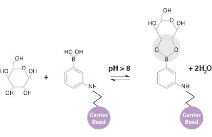
Glycoprotein Enrichment Resin是一种硼酸基树脂,能够从复杂样品(例如人血清)中快速、有效、特异性地富集糖基化蛋白质,可以使用简单的重力流方法或中等压力的方法(例如FPLC)进行操作。树脂是偶联了间氨基苯硼酸配体的琼脂糖珠,该配体能够结合存在于糖蛋白分子糖部分内的残基如甘露糖、半乳糖或葡萄糖,与这些糖残基的顺式二醇基团形成可逆的五元环络合物。这种复合物可通过降低pH,或使用含Tris或山梨糖醇的洗脱buffer使其分离。
 
■ 产品特点
灵活、有效、特异性的富集和检测糖基化蛋白质。
· 灵活——使用本树脂可以应用重力流法或FPLC法
· 性能出色——本树脂特异性结合糖蛋白能力更强,而非特异性结合很弱
· 特异性——本树脂对血清中的高、低丰度糖蛋白均能够有效富集
· 快速——90 min内即可完成糖蛋白的富集
· Western Detection Kit可提供快速(1 hr)、灵敏、特异性的不依赖抗体的检测
 
■ 应用
糖蛋白的富集和检测
 
■ 实验例
 
糖蛋白富集 & 检测
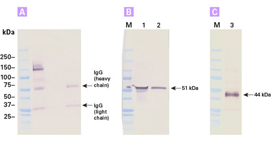
通过Western Blotting对Glycoprotein Enrichment Resin纯化产物进行分析,结果显示,该树脂特异性地富集了高丰度和低丰度的血清糖蛋白。图A.使用IgG抗体检测到特异性富集的高丰度血清糖蛋白IgG,lane 1是人血清,lane 2是流穿液,lane 3是洗脱液(糖蛋白)。图B和C是通过Western Blotting对纯化得到的两种特异性的低丰度血清糖蛋白α-1酸性糖蛋白(图B)和α-1抗胰蛋白酶(图C)进行检测的结果,lane 1是人血清,lane 2是洗脱组分,lane 3是洗脱组分。


产品详情请点击:糖蛋白富集 & 检测


页面更新:2019-11-12 10:52:12

49006010-东洋直径10mm抗微生物物质纸盘

  • 型号 49006010
  • 品牌 ADVANTEC东洋
  • 【简单介绍】
    品牌 其他品牌 抗生素物质纸盘 直径10mm

    东洋直径10mm抗微生物物质纸盘,抗生素分析检测抗菌物质检测肉类青*素纸盘 纯纤维素素纤维制造 高吸收效能三种类型可以用于以下用途 抗生素分析 检测试题抗菌物质检测肉类青*素

    东洋直径10mm抗微生物物质纸盘

    ADVANTEC TOYO抗生素分析检测抗菌物质检测肉类青*素纸盘49006010

    抗生素分析检测抗菌物质检测肉类青*素纸盘 纯纤维素素纤维制造 高吸收效能三种类型可以用于以下用途 抗生素分析 检测试题抗菌物质检测肉类青*素

    抗生素分析检测抗菌物质检测肉类青*素纸盘 纯纤维素素纤维制造 高吸收效能三种类型可以用于以下用途 抗生素分析 检测试题抗菌物质检测肉类青*素。

    东洋直径10mm抗微生物物质纸盘

    49006010-东洋直径10mm抗微生物物质纸盘


Lumitester C-110 ATP荧光检测仪C-110 品牌:Kikkoman


品牌:Kikkoman
CAS No.:
储存条件:室温
纯度:
产品编号

(生产商编号)

等级 规格 运输包装 零售价(RMB) 库存情况 参考值

301-34671

1 EA

Lumitester C-110                                                      ATP荧光检测仪C-110            品牌:Kikkoman

替代品:61911


* 干冰运输、大包装及大批量的产品需酌情添加运输费用


* 零售价、促销产品折扣、运输费用、库存情况、产品及包装规格可能因各种原因有所变动,恕不另行通知,确切详情请联系上海金畔生物科技有限公司。

产品描述相关资料下载相关产品浏览记录

运用试剂,检测ATP浓度,微生物情况,检测时间约10秒。
主要应用:
1. 利用C-110,通过检测一系列不同浓度菌数(CFU)的RLU,制作成标准曲线。以后对照曲线图,检测RLU,推测对应的CFU。
2. 检测有无菌
3. 检测是否有大肠杆菌
4. 检测ATP量
可用的试剂有两类:
一、LuciPacII  (试剂棒,操作方法与PD-30配套试剂LuciPac Pen相同,用来检测ATP量)
二、Lucifell/CheckLite系列(试剂盒,检测微生物量、大肠菌群、有无菌等)
Lucifell 250 Plus/CheckLite 250 Plus:ATP浓度・微生物量的检测
Lucifell HS SET/CheckLite HS SET:更灵敏测出ATP浓度,微生物量
Lucifell AT100/CheckLite  AT100 :无菌实验等各种微生物检查
Lucifell CT150/CheckLite CT150 :检测出大肠菌群(根据样品增菌速度,最短5.5 小时)
Lucifell ATP Eliminating Reagent Set/CheckLite Eliminating Reagent Set :去除试剂盒  去除微生物以外 ATP
Lucifell ATP Standard Reagent Set/CheckLite Standard Reagent Set:标准试剂盒  用于制作发光量的校准线

1226556-GVS 重复检测带电尼龙转印膜孔径0.45um卷膜

  • 型号 1226556
  • 品牌 GVS
  • 【简单介绍】

    GVS 重复检测带电尼龙转印膜孔径0.45um卷膜 1226556
    Maine Manufacturing重复检测带电尼龙转印膜是一种投到惰性聚酯网的纯聚合物。它具有天然亲水性和固有带电,使其非常适合多次重复检测并用于碱性条件下的核酸转移。


    简介和用途
    Maine Manufacturing重复检测带电尼龙转印膜是一种投到惰性聚酯网的纯聚合物。它具有天然亲水性和固有带电,使其非常适合多次重复检测并用于碱性条件下的核酸转移。
    典型应用
      .Southern转移
      .Northern转移
      .碱性转移
      .放射性标记检测系统
      .多次重复检测
      .微阵列
      .巨阵列
      .化学发光系统
    特点和优点
     Maine Manufacturing重复检测带电尼龙转印膜是一种受到支撑的带电尼龙膜。
    出众的结合能力,可靠的结果
     Maine Manufacturing重复检测带电尼龙转印膜专门针对碱性条件下和多次再检测时的核酸转移而设计。它也可以被用于放射性和非放射性探针,包括化学发光系统和生物化检测系统。
    鉴于较小的样品和更高密度形式或微阵列变得越来越普遍, Maine Manufacturing重复检测带电尼龙转印膜尤其适合于基因组学研究。

    GVS 重复检测带电尼龙转印膜孔径0.45um卷膜 1226556

    1226556-GVS 重复检测带电尼龙转印膜孔径0.45um卷膜

49005040-ADVANTEC东洋抗生素活性检测纸6mm

  • 型号 49005040
  • 品牌 ADVANTEC东洋
  • 【简单介绍】

    ADVANTEC东洋抗生素活性检测纸6mm抗生素分析检测抗菌物质检测肉类青霉素纸盘

    ADVANTEC东洋抗生素活性检测纸6mm

    抗生素分析检测抗菌物质检测肉类青霉素纸盘 纯纤维素素纤维制造 高吸收效能三种类型可以用于以下用途 抗生素分析 检测试题抗菌物质检测肉类青霉素

    ADVANTEC东洋抗生素活性检测纸6mm


49006010-ADVANTEC抗微生物物质纸盘分析纸PAPER DISC

  • 型号 49006010
  • 品牌 ADVANTEC东洋
  • 【简单介绍】

    ADVANTEC抗微生物物质纸盘分析纸PAPER DISC抗生素分析检测抗菌物质检测肉类青霉素纸盘 纯纤维素素纤维制造 高吸收效能三种类型可以用于以下用途 抗生素分析 检测试题抗菌物质检测肉类青霉素


    49006010-ADVANTEC抗微生物物质纸盘分析纸PAPER DISC

药典检测用溶剂 符合日本药典、美国药典和欧洲药典用于药物检测的试剂

  • 产品特性
  • 相关资料
  • Q&A
  • 参考文献

药典检测用溶剂药典检测用溶剂                              符合日本药典、美国药典和欧洲药典用于药物检测的试剂

符合日本药典、美国药典、欧洲药典

  这些用于药物检测的试剂符合三大药典的要求,即日本药典、美国药典和欧洲药典。目前,Wako 已发布了多种用于普通药物检测的试剂。


◆ 认证体系

● 日本药典(JIS special grade reagent)

● 美国药典(ACS Specification)

● 欧洲药典

◆认证检测项目 (例: 丙酮)

检测项目

日本药典
(JIS Special Grade)

美国药典(ACS)

欧洲药典

纯度(丙酮)

99.5+%(质量含量)

99.5+%

外观

无色透明液体*

颜色(APHA)(USP-NF方法)

≤10

不挥发物残余
 (蒸发残渣)(EP方法)

≤5 ppm(质量含量)

≤0.001%

≤50 ppm

水中溶解性(水不溶性物质)(USP-NF 方法)

通过

通过

通过

可滴定酸(USP-NF 方法)

≤0.002%(质量含量)

≤ 0.0003 meq/g

通过

可滴定碱(USP-NF方法)

≤0.001%(质量含量)

≤ 0.0006 meq/g

通过

醛类(以甲醛计)(USP-NF方法)

≤0.002%(质量含量)

≤ 0.002%

异丙醇(USP-NF方法)

M≤ 0.05%

甲醇(USP-NF method)

≤0.05%(质量含量)

≤ 0.05%

相关物质(EP方法)

通过

高锰酸盐还原性化合物 
(USP-NF方法)

≤ 2%(质量含量)

通过

通过

水(H2O)(USP-NF方法)

≤0.3%(质量含量)

≤0.5

≤3 g/L

折射率 nD20

1.358-1.360

密度(相对)

0.789-0.792g/mL

0.790~0.793


 * 表示 Wako 厂家自己添加的检测项目

◆ 产品列表

产品编号

产品名称

认证体系

包装

级别

012-23325

Acetic Acid

醋酸

JIS, ACS, Reag. EP

500 mL

for Medicine Test

019-23335

Acetic Acid Anhydride

醋酸酰

JIS, ACS, Reag. EP

500 mL

016-23345

Acetone

丙酮

JIS, ACS, Reag. EP

500 mL

013-23355

Ammonia Solution

氨溶液

JIS, ACS, Reag. EP

500 mL

044-31615

Diammonium Hydrogen Citrate

柠檬酸氢二铵

JIS, ACS, Reag. EP

500 g

047-31605

Diethyl Ether*

二乙醚

JIS, Reag. EP

500 mL

048-31635

N,N-Dimethylformamide

N,N-二甲基甲酰胺

JIS, ACS, Reag. EP

500 mL

041-31505

Disodium Hydrogenphosphate 12-Water

磷酸氢二钠12水合物

JIS, Reag. USP, Reag. EP

500 g

053-08155

Ethyl Acetate

乙酸乙酯

JIS, ACS, Reag. EP

500 mL

055-08095

Ethanol (95)

乙醇(95)

JIS, Reag. USP, Reag. EP

500 mL

058-08085

Ethanol (99.5)

乙醇(99.5)

JIS, ACS, Reag. EP

500 mL

084-09205

Hydrochloric Acid*

盐酸

JIS, Reag. EP

500 mL

130-16585

Methanol*

甲醇

JIS, Reag. EP

500 mL

160-24795

2-Propanol*

异丙醇

JIS, Reag. EP

500 mL

167-24685

Potassium Dihydrogen Phosphate 

磷酸二氢钾

JIS, ACS, Reag. EP 500 g

198-15965

Sodium Acetate

乙酸钠

JIS, ACS, Reag. EP

500 g

198-16065

Sodium Acetate Trihydrate

乙酸钠二水合物

JIS, ACS, Reag. EP

500 g

195-15975

Sodium Chloride

氯化钠

JIS, ACS, Reag. EP

500 g

192-15985

Sodium Hydrate

氢氧化钠

JIS, ACS, Reag. EP

500 g

199-15995

Sulfuric Acid*

硫酸

JIS, Reag. EP

500 mL

203-18465

Toluene

甲苯

JIS, ACS, Reag. EP

500 mL


 *表示符合两大药典(日本药典和欧洲药典)。 Wako 还可以提供符合美国药典的检测结果(仅有部分检测项目)。

◆相关产品

符合药典要求的液相色谱用溶剂

以下产品是符合日本药典、美国药典、欧洲药典要求的溶剂 

产品编号

产品名称

包装

级别

019-21691

Acetonitrile

乙腈

1 L

日本药典(液相色谱)

015-21693

3 L

136-15661

Methanol

甲醇

1 L

132-15663

3 L

085-08711

Hexane

己烷

1 L

081-08713

3 L

产品编号 产品名称 产品规格 产品等级

残留溶剂检测用溶剂 用于药品的残留溶剂检测

  • 产品特性
  • 相关资料
  • Q&A
  • 参考文献

用于药品的残留溶剂检测残留溶剂检测用溶剂                              用于药品的残留溶剂检测

残留溶剂检测用溶剂

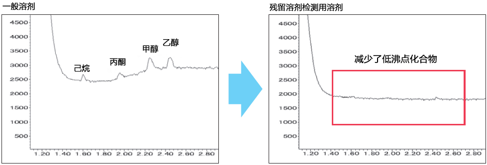
药品中的残留溶剂在《药品残留溶剂指南》(ICH Q3C)中,按照影响人体健康的风险程度,被归入Class 1~3管理。目前日本药典和美国药典(USP)对残留药物溶剂的检测方法中,规定使用顶空气相色谱法的高灵敏度分析方法,但部分市售溶剂可能存在其他杂质溶剂的残留,当溶剂峰被检测到时,无法判断杂质究竟来自样品还是来自溶剂,导致无法获得正确的检测结果。

FUJIFILM Wako的“残留溶剂检测用”溶剂,减少了溶剂的低沸点化合物,并保证其含量处于低水平。本产品系列共有两种类型产品:一种是保证收录于ICH Q3C Class 1~3(ICH Q3C检测用)残留溶剂含量低的检测用溶剂;另一种是保证FUJIFILM Wako选定的4种溶剂的低残留溶剂。


◆残留溶剂检测用(ICH Q3C检测用)


√ 溶剂中含有的低沸点化合物得到减少!

残留溶剂检测用溶剂                              用于药品的残留溶剂检测

√ 保证低含量:针对收录于ICH Q3C Class 1~3的59种残留溶剂


除残留溶剂检测用(见背面)的溶剂外,还保证收录于Class 1~3中的以下溶剂类型的残留量低。


Class 1(低于1 ppm)

四氯化碳

1,2-二氯乙烷

1,1-二氯乙烯

1,1,1-三氯乙烷

Class 2(低于10 ppm)※1

乙腈

乙苯

2-乙氧基乙醇

邻、间、对二甲苯

氯苯

异丙苯 ※2

氯仿

环己烷

1.,2-二氯乙烯

1,4-二恶烷

二氯甲烷

N,N-二甲基乙酰胺

N,N-二甲基甲酰胺

1,2-二甲氧基乙烷

环丁砜

四氢呋喃 ※2

四氢萘

甲苯

1,1,2-三氯乙烯

硝基甲烷

吡啶

己烷

甲醇

甲基异丁基酮 ※2

甲基环己烷

N-甲基吡咯烷酮

甲基丁基酮

2-甲氧基乙醇

※1 不保证乙二醇和甲酰胺的含量。

※2 保证低于50 ppm。

Class 3(低于50 ppm)

丙酮

苯甲醚

乙醇

甲酸乙酯

醋酸

乙酸异丁酯

醋酸异丙酯

乙酸乙酯

乙酸正丁酯

醋酸丙酯

醋酸甲酯

乙醚

二甲基亚砜

1-丁醇

2-丁醇

叔丁基甲基醚

1-丙醇

2-丙醇

庚烷

1-戊醇

戊烷

甲乙酮

3-甲基-1-丁醇

2-甲基-1-丙醇

※ 不保证甲酸和三乙胺的含量。

产品编号

产品名称

规格

包装

049-34725

1,3-二甲基-2-咪唑烷酮(DMI) ICH Q3C检测用

残留溶剂检测用

500 mL

◆残留溶剂检测用


溶剂中的低沸点化合物得到减少,保证以下4种溶剂类型低于0.1 ppm。


保证溶剂类型(低于0.1 ppm)

丙酮

乙醇

甲醇

己烷

※  保证溶剂的类型不同于残留溶剂检测用溶剂(ICH Q3C检测用)

产品编号

产品名称

规格

包装

043-31565

N,N-Dimethylacetamide

N,N-二甲基乙酰胺

残留溶剂检测用

500 mL

041-31561

3 L

044-33315

1,3-Dimethyl-2-imidazolidinone

1,3-二甲基-2-咪唑啉酮

残留溶剂检测用

500 mL

042-33311

3 L

046-31555

N,N-Dimethylformamide

N,N-二甲基甲酰胺(DMF)

残留溶剂检测用

500 mL

044-31551

3 L

041-31365

Dimethyl Sulfoxide

二甲基亚砜(DMSO)

残留溶剂检测用

500 mL

049-31361

3 L

◆药物中的杂质管理


残留溶剂检测用溶剂                              用于药品的残留溶剂检测

※ 本页面产品仅供研究用,研究以外不可使用。

产品编号 产品名称 产品规格 产品等级

PROTEOSTAT® PDI assay kit 蛋白二硫键异构酶检测试剂盒

  • 产品特性
  • 相关资料
  • Q&A
  • 参考文献

PROTEOSTAT® PDI assay kitPROTEOSTAT®  PDI assay kit                              蛋白二硫键异构酶检测试剂盒

蛋白二硫键异构酶检测试剂盒

PROTEOSTAT® PDI 检测试剂盒能够以稳定且高通量的方式定量检测PDI酶活性,适用于从化学库中筛选PDI抑制剂,为筛选PDI酶活性调节剂提供了一种简单、均相的检测方法。

本试剂盒在二硫苏糖醇(DTT)存在的条件下,PDI酶催化胰岛素聚集体的形成,随后与发射红色荧光的 PROTEOSTAT® PDI检测染料结合,通过监测胰岛素减少来实现对PDI酶活性的检测。

相对于使用比浊法检测PDI酶活性,基于荧光的检测法有着极大改进的信号检测窗口和检测下限,以及优越的 Z' 因子(>0.8)。检测中板内和板间CV值为3-5%。

本试剂盒中包含人重组PDI酶(EC 5.3.4.1)、杆菌肽(PDI抑制剂)以及监测PDI酶活性变化所需的全部试剂。

◆产品详情


基础信息


别称:蛋白二硫键异构酶(Protein disulfide isomerase)检测试剂盒

应用:荧光检测

储存条件:避光。避免反复冻融。

运输方式:干冰运输

长期储存温度:–80°C

特点:本试剂盒为筛选蛋白二硫键异构酶(PDI)酶活性的调节剂提供了一种简单、均相的检测方法。

UniProt ID:P07237(人)

监管状态:RUO – 仅供研究使用

技术信息/产品说明:Enzo和PROTEOSTAT®是Enzo Life Sciences, Inc. 的商标。

技术信息/产品说明:Enzo的部分产品和产品应用受美国、外国专利及正在申请的专利保护。


品质保证


(1)按照手册中的步骤, PROTEOSTAT® PDI检测试剂盒可用于检测以下样品:

(1)① 含PDI的胰岛素;② 不含PDI的胰岛素;③ 含有PDI和杆菌肽的胰岛素。

(2)Z' 因子高于0.8。

(3)PDI活性至少可被杆菌肽抑制50%。


使用/稳定性


在产品标签注明的日期前,试剂盒组分在适当的储存条件下可稳定保存。

可存放于-20°C的无霜冰箱中,或在–80°C下长期保存。


组分


● PROTEOSTAT® PDI 检测试剂

● PDI(人)(重组)

● 胰岛素(冻干)

● 杆菌肽(抑制剂对照,冻干)

● PBE 缓冲液

● 终止试剂

● DTT(10 mM)

● 去离子水


※ 本页面产品仅供研究用,研究以外不可使用。

参考文献


 1. 

Thioredoxin reductase is a major regulator of metabolism in leukemia cells: S. Karunanithi, et al.; Oncogene 41388 (2021), Abstract;


 2.

Molecular docking‐assisted screening reveals tannic acid as a natural protein disulphide isomerase inhibitor with antiplatelet and antithrombotic activities: L. Ren, et al.; J. Cell. Mol. Med. 24, 14257 (2020), AbstractFull Text

 3.

PDI knockdown inhibits seizure activity in acute seizure and chronic epilepsy rat models via S-nitrosylation-independent thiolation on NMDA receptor: A.R. Jeon, et al.; Front. Cell. Neurosci. 12, 438 (2018), Application(s): Hippocampal tissue lysates, AbstractFull Text

 4.

The thiosemicarbazone Me2NNMe2 induces paraptosis by disrupting the ER thiol redox homeostasis based on protein disulfide isomerase inhibition: S. Hager, et al.; Cell Death Dis. 9, 1052 (2018), AbstractFull Text

 5.

Tissue Factor Coagulant Activity is Regulated by the Plasma Membrane Microenvironment: Y. Yu, et al.; Thromb. Haemost. 118, 990 (2018), Application(s): In-vitro activity assay, Abstract;


 6.

Curcumin and Curcuma longa L. extract ameliorate lipid accumulation through the regulation of the endoplasmic reticulum redox and ER stress: H.Y. Lee, et al.; Sci. Rep. 7, 6513 (2017), Application(s): PDI assay of tissue lysates, AbstractFull Text

 7.

Nox4 regulates the eNOS uncoupling process in aging endothelial cells: H.Y. Lee, et al.; Free Radic. Biol. Med. 113, 26 (2017), Application(s): PDI assay of HUVEC cell lysates, Abstract;


 8.

PDI regulates seizure activity via NMDA receptor redox in rats: J.Y. Kim, et al.; Sci. Rep. 7, 42491 (2017), Application(s): Hippocampal tissue lysates, Abstract; Full Text

 9.

Protein disulfide isomerase regulates renal AT1 receptor function and blood pressure in rats: X. Wang, et al.; Am. J. Physiol. Renal. Physiol. 313, F461 (2017), Application(s): Renal tissue lysates, AbstractFull Text

10.

Requirement of zinc transporter SLC39A7/ZIP7 for dermal development to fine-tune endoplasmic reticulum function by regulating protein disulfide isomerase: B.H. Bin, et al.; J. Invest. Dermatol. 137, 1682 (2017), Application(s): ER fractions from murine osteoblasts, Abstract;


11. 

Succination of protein disulfide isomerase links mitochondrial stress and endoplasmic reticulum stress in the adipocyte during diabetes: A.M. Manuel, et al.; Antioxid. Redox Signal. 27, 1281 (2017), Application(s): 3T3-L1 adipocytes and adipose tissue lysates, AbstractFull Text

12.

Adverse outcomes associated with cigarette smoke radicals related to damage to protein disulfide isomerase: H. Kenche, et al.; J. Biol. Chem. 291, 4763 (2016), Abstract;


13.

Adverse Outcomes Associated with Cigarette Smoke Radicals Related to Damage to Protein-disulfide Isomerase: H. Kenche, et al.; J. Biol. Chem. 291, 4763 (2016), Application(s): in-vitro assay with recombinant PDI, AbstractFull Text

14.

Role of two sequence motifs of mesencephalic astrocyte-derived neurotrophic factor in its survival-promoting activity: K. Mätlik, et al.; Cell Death Dis. 6, e2032 (2015), AbstractFull Text

15.

Methods of measuring protein disulfide isomerase activity: a critical overview: M.M. Watanabe, et al.; Front. Chem. 2, 73 (2014), Application(s): Review on available assays to measure PDI activity, AbstractFull Text

16.

N-octanoyl dopamine treatment of endothelial cells induces the unfolded protein response and results in hypometabolism and tolerance to hypothermia: E. Stamellou, et al.; PLoS One 9, e99298 (2014), AbstractFull Text

17.

The protein disulfide isomerases PDIA4 and PDIA6 mediate resistance to cisplatin-induced cell death in lung adenocarcinoma: G. Tufo, et al.; Cell Death Differ. 21, 685 (2014), Application(s): Evaluation of PDI activity in whole extracts and ER fractions, Abstract;


18.

Rapid activation of monocyte tissue factor by antithymocyte globulin is dependent on complement and protein disulfide isomerase: F. Langer, et al.; Blood 121, 2324 (2013), Abstract;


19.

Oxidative processing of latent Fas in the endoplasmic reticulum controls the strength of apoptosis: V. Anathy, et al.; Mol. Cell. Biol. 32, 3464 (2012), Abstract;


◆一般参考文献


 1. 

Attachment and entry of Chlamydia have distinct requirements for host protein disulfide isomerase: S. Abromaitis & R.S. Stephens; PLoS Pathog. 5, e1000357 (2009), Abstract;


 2.

Protein disulfide isomerase (PDI) associates with NADPH oxidase and is required for phagocytosis of Leishmania chagasi promastigotes by macrophages: C. X. Santos, et al.; J. Leukoc. Biol. 86, 989 (2009), Abstract;


 3.

The proteostasis boundary in misfolding diseases of membrane traffic: D.M. Hutt, et al.; FEBS Lett. 583, 2639 (2009), Abstract;


 4.

Increasing melanoma cell death using inhibitors of protein disulfide isomerases to abrogate survival responses to endoplasmic reticulum stress: P.E. Lovat, et al.; Cancer Res. 68, 5363 (2008), Abstract;


 5.

Targeting homeostatic mechanisms of endoplasmic reticulum stress to increase susceptibility of cancer cells to fenretinide-induced apoptosis: the role of stress proteins ERdj5 and ERp57: M. Corazzari, et al.; Br. J. Cancer 96, 1062 (2007), Abstract;


 6.

Coupling endoplasmic reticulum stress to the cell death program: R.V. Rao, et al.; Cell Death Differ. 11, 372 (2004), Abstract;


 7.

Protein-disulfide isomerase-mediated reduction of two disulfide bonds of HIV envelope glycoprotein 120 occurs post-CXCR4 binding and is required for fusion: R. Barbouche, et al.; J. Biol. Chem. 278, 3131 (2003), Abstract;


 8.

Protein disulfide isomerase, a component of the estrogen receptor complex, is associated with Chlamydia trachomatis serovar E attached to human endometrial epithelial cells: C.H. Davis, et al.; Infect. Immun. 70, 3413 (2002), Abstract;


 9.

Protein-disulfide isomerase-mediated reduction of the A subunit of cholera toxin in a human intestinal cell line: P.A. Orlandi; J. Biol. Chem. 272, 4591 (1997), Abstract;


10.

Acceleration of reactivation of reduced bovine pancreatic ribonuclease by a microsomal system from rat liver: R.F. Goldberger, et al.; J. Biol. Chem. 238, 628 (1963), Abstract;


产品编号 产品名称 产品规格 产品等级
ENZ-51024-KP002 PROTEOSTAT PDI assay kit 
PROTEOSTAT PDI检测试剂盒
2×96 tests
ENZ-51024-KP050 PROTEOSTAT PDI assay kit 
PROTEOSTAT PDI检测试剂盒
50 tests

Adiponectin (human) ELISA Kit 脂联素 (人) ELISA试剂盒 品牌:Adipogen


品牌:Adipogen
CAS No.:
储存条件:+4°C
纯度:
产品编号

(生产商编号)

等级 规格 运输包装 零售价(RMB) 库存情况 参考值

AG-45A-0001YEK-KI01

96 wells 5,160.00


* 干冰运输、大包装及大批量的产品需酌情添加运输费用


* 零售价、促销产品折扣、运输费用、库存情况、产品及包装规格可能因各种原因有所变动,恕不另行通知,确切详情请联系上海金畔生物科技有限公司。

产品描述相关资料下载相关产品浏览记录 代谢研究相关试剂。
检测物种:人。
检测灵敏度:100pg/mL。
检测范围:0.5~32ng/mL。
样品类型:细胞培养上清,血清,血浆。
检测类型:夹心法。

Adiponectin (human) ELISA Kit (Twin Plex) 脂联素 (人) ELISA试剂盒 (2盒) 品牌:Adipogen


品牌:Adipogen
CAS No.:
储存条件:+4°C
纯度:
产品编号

(生产商编号)

等级 规格 运输包装 零售价(RMB) 库存情况 参考值

AG-45A-0001YTP-KI01

2 x 96 wells 9,000.00


* 干冰运输、大包装及大批量的产品需酌情添加运输费用


* 零售价、促销产品折扣、运输费用、库存情况、产品及包装规格可能因各种原因有所变动,恕不另行通知,确切详情请联系上海金畔生物科技有限公司。

产品描述相关资料下载相关产品浏览记录 代谢研究相关试剂。
检测物种:人。
检测灵敏度:100pg/mL。
检测范围:0.5~32ng/mL。
样品类型:细胞培养上清,血清,血浆。
检测类型:夹心法。

骨吸收检测试剂盒 Bone Resorption Assay Kit

  • 产品特性
  • 相关资料
  • Q&A
  • 参考文献

骨吸收检测试剂盒骨吸收检测试剂盒                              Bone Resorption Assay Kit

Bone Resorption Assay Kit


◆原理


骨吸收检测试剂盒                              Bone Resorption Assay Kit

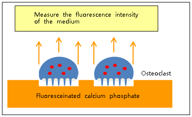
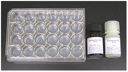
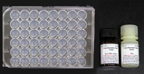
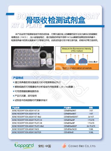
       本产品是用于检测骨吸收作用的试剂盒,已预先铺在板上的磷酸钙首先与荧光素标记的硫酸软骨素结合(FACS),加入破骨细胞后,通过细胞的呼吸作用将 FACS 从磷酸钙层释放到培养基中,检测培养基中的荧光强度来评价骨吸收作用。此款试剂盒可用于骨生理代谢、药物评价等方面研究。


骨吸收检测试剂盒                              Bone Resorption Assay Kit

                BONE RESORPTION ASSAY KIT 24

骨吸收检测试剂盒                              Bone Resorption Assay Kit

                   BONE RESORPTION ASSAY KIT 48

骨吸收检测试剂盒                              Bone Resorption Assay Kit

骨吸收检测试剂盒                              Bone Resorption Assay Kit


◆优点特色


● 简单快捷——仅需检测荧光强度变化即可评估骨吸收能力

● 操作方便——观察骨吸收后形成的凹洞,仅需移除细胞。

● 准确清晰——可透过显微镜观察细胞形态,并可用图像软件分析吸收效果。

● 创新有效——使用标定荧光的磷酸钙来取代传统的牙质骨片。

● 产品已灭菌,即可使用


◆案例应用


<药物作用巨噬细胞 RAW264 细胞系后实验检测>


1. 剂盒的板里每孔加入 FACS,37℃ 孵育1-2小时。


2. PBS 和培养基漂洗后,加入巨噬细胞 RAW264 细胞

    孵育。培养的细胞已用细胞分化诱导剂。

  (RANKL)和药物处理过(约培养5-6天)。


3. 养基转到 96 孔酶标板中,加入 Assay Buffer。

    在入射光 485 nm、发射光 535 nm 的条件下进行荧

    强度检测。


4. 分析吸收作用效果图,使用5%次氯酸钠处理可移走

    胞,经过漂洗后,用显微镜拍照,最后用图像分析

    分析吸收作用效果图。

骨吸收检测试剂盒                              Bone Resorption Assay Kit

注意:(1)使用无酚红培养基。(2)使用时请避光。

检测试剂盒

产品编号

名称

应用

规格

储存

PMC-AK03-COS

Acidic Mucopolysaccaride Assay Kit

定量检测酸性粘多糖

1 kit (100 tests)

4-10℃
勿冻

试剂盒组成

● Chromogenic Solution (4 mL) ——1  bottle 

● Buffer (130 mL) ——1 bottle                  

● Enzyme Powder For Sample Preparation ——5 bottles

● Chondroitin Sulfate Standard Solution (100 µg/mL)(2 mL) —— 1 bottle

PMC-AK20-COS

Alkaline Phosphatase Staining Kit

检测成骨细胞碱性磷酸酶

1 kit (for 12 well plate×10)

4-10℃
勿冻

试剂盒组成

● Chromogenic Substrate ——10 vials                    

● Substrate-containing Buffer (50 mL) ——1 bottle

PMC-AK21-COS

Calcified Nodule Staining Kit

检测成骨细胞钙化结节

1 kit (for 24 well plate×10)

RT

试剂盒组成

● Chromogenic Substrate —— 10 vials                  

● Substrate-containing Buffer (×100)  (60 mL) —— 1 bottle

PMC-AK04F-COS

TRAP Staining Kit
TRAP: 抗酒石酸酸性磷酸酶

检测破骨细胞抗酒 石酸酸性磷酸酶

1 kit(for 24 well plate×10)

4-10℃
勿冻

试剂盒组成

● Chromogenic Substrate——10 vials 每个试剂盒所包含试剂可用于10×96-孔板

● Tartrate-containing Buffer (50 mL)——1 bottle

294-67001

TRAP/ALP Stain Kit

骨代谢研究

60回用

-20℃

PMC-AK02-COS

Stains All Gel Staining Kit

酸性蛋白染色

1 kit (20 test)

RT

 特色:

   酸性蛋白调节骨骼钙化,如骨钙素、骨桥蛋白和 BSP,这些蛋白是骨骼和牙齿的重要成分。但这些酸性蛋白难以通过传统的 SDS-PAGE 凝胶染色来检测。Stains All Gel Staining Kit 是针对这些难染色的强酸性蛋白设计,蛋白条带的颜色取决于蛋白的等电点和化学修饰,如糖基化和磷酸化。

试剂盒组成

● Staining Stock Solution ( ×10) ——40 mL

● Dilution Buffer —— 200 mL×2

CSR-ARD-SET

Calcification Evaluation Set

检测成骨细胞钙化

1 SET

RT

试剂盒组成

● Alizarin Red Solution (CSR-ARD-A1) ——100 mL

● Calcified Nodule Extraction Solution(CSR-ARD-E1) ——100 mL


其他

产品编号

名称

检测样本

规格

储存

291-58601

LabAssay ALP

小鼠血清等

900 test

2-10℃勿冻

ALX-850-019-KI01

RANKL (total), soluble  (human) ELISA kit

人血清、血浆、细胞培养液

96 well

 4°C

APO-54N-028-KI01

Osteoprotegerin free (human), detection set

人血清、血浆、细胞培养液

96 well

 4°C

ALX-850-280A-KI01

Osteoprotegerin (human)  ELISA kit

人血清、

血浆

96 well

 4°C

ADI-900-215-0001

25(OH) Vitamin D ELISA kit

人血清、

血浆

96 well

 -20℃

骨吸收检测试剂盒                              Bone Resorption Assay Kit

骨吸收检测试剂盒                              Bone Resorption Assay Kit骨吸收检测试剂盒

骨吸收检测试剂盒                              Bone Resorption Assay Kit

骨吸收检测试剂盒                              Bone Resorption Assay KitDATA SHEET.pdf

骨吸收检测试剂盒                              Bone Resorption Assay Kit

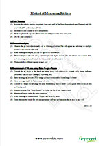
骨吸收检测试剂盒                              Bone Resorption Assay KitMethod of Measuring Pit Area.pdf

骨吸收检测试剂盒                              Bone Resorption Assay Kit

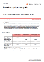
骨吸收检测试剂盒                              Bone Resorption Assay KitBRA24KIT_E.20150529.pdf

骨吸收检测板/试剂盒【常见问题】

Q1:    培养基的使用量?
A1:    BONE RESORPTION ASSAY KIT 48×2(CSR-BRA-48X2KIT)按照说明书,该试剂盒需要用分化培养基处理板子。1个板子,48孔,
            每个孔需用 0.5 mL 分化培养基处理,每个孔加入细胞(细胞含有 0.5 mL分化培养基)。1个48孔板合计需要的分化培养基是48 mL。

            2个48孔板,是 96 mL分化培养基。


           Osteoclast Culture Kit V-2 (PMC-OSC12-COS )这个试剂盒中含有25 mL的分化培养基。所以,Osteoclast Culture Kit V-2 

         (PMC-OSC12-COS )里面的分化培养基25 mL是远远不够用于BONE RESORPTION ASSAY KIT 48×2(CSR-BRA-48X2KIT)的检测。


           Osteoclast Culture Kit V-2 (PMC-OSC12-COS )即便里面的分化培养基不含酚红,也需要另外配置。

Q2:    如何配置不含酚红的培养基?
A2:   不含酚红的分化培养基配方:
       1)alpha-MEM(不含酚红),10%胎牛血清,1%双抗,50 ng/mL M-GSF,15 ng/mL RANKL。Osteoclast Culture Kit V-2 中用的也是

            alpha-MEM。
       2)DMEM/F12(不含酚红),10%胎牛血清,1%双抗,50 ng/mL M-GSF,15 ng/mL RANKL。Osteoclast Culture Kit V-2 中用的也是

            alpha-MEM不同。BONE RESORPTION ASSAY KIT 48×2 (CSR-BRA-48X2KIT)试剂盒中用到的。

Q3    在细胞孵育(5~6天)后,最佳的荧光检测时间是什么?什么时候荧光强度开始减弱? 什么时候必须检测荧光强度?

A3:    最佳荧光检测时间取决于细胞和培养状态。所以我们推荐继续使用少量的条件培养基,研究荧光强度的上升曲线。


Q4:    板的保质期多久?

A4:    距离发货时间1年。


Q5:    板的荧光强度会随时间而减弱吗?

A5:    避光情况下,结合磷酸钙(CaP)的荧光标记硫酸软骨素(FACS)的荧光强度稳定。然而,部分荧光标记硫酸软骨素(FACS)在有培养

     基或者PBS时会被洗脱到培养基内。


Q6:    如果我用胰酶消化细胞做 qPCR,磷酸钙(Ca-P)是否影响胰酶消化或者 qPCR 结果?

A6:    非常抱歉,我们没有实验过,也没有相关数据。


Q7:    我如何检测 pit?与 TRAP 检测类似吗?需要检测 pit 的深度和宽度吗?(检测 pit 的面积不等于检测破骨细胞消化的 pit 体积)

A7:     Pit 检测(骨吸收活性)与 TRAP 检测不是很相似。按照说明书,pit 检测中细胞的面积是在移除细胞后对板进行拍照测量。实际上,如您

             所说体积不应该在 pit 检测中测量。检测体积也比较困难。因此,pit 面积或者 pit 数目经常检测。


Q8:    我该如何洗涤细胞?用缓冲液?检测荧光强度的 pit 实验后。

A8:    用5%的次氯酸钠处理5分钟将细胞从板上移除,然后用水洗涤板,晾干。


Q9:    可用 TECAN 的活细胞影像仪器对板进行实时检测?

A9:    通常,我们会在新的 96 孔板内收集条件培养基,检测荧光强度。我们没有在培养过程中检测荧光强度。


Q10:    根据说明书,需要使用非酚红培养基培养细胞,我是否可以用其他不含酚红的培养基(不是说明书上提到的培养基)?

              这种培养基是否对细胞有不利影响?能否推荐其他不含酚红的培养基。我比较担心在磷酸钙(CaP)包被的培养板上培养细胞,不含酚红

              的 DMEM 或者其他培养基不能很好的支持细胞培养5天。在将细胞接种在磷酸钙(CaP)包被的培养板之前,我是否可以用含酚红的培

              基培养细胞?我想培养 RAW264.7 细胞和 U937 细胞。另外,5%的次氯酸钠是否也包含在试剂盒里?

A10:    在我们培养体系中,不更换培养基可以培养6天。如果客户担心6天培养的问题,我们推荐他试试在正常培养板中进行同样的培养,确认

              否可以诱导破骨细胞。

              非常抱歉,我们这边没有用除了 DMEM/F-12、αMEM 之外的培养基。可以试试,确认是否能诱导破骨细胞。在最初培养的时候,可以

              含酚红的培养基。但在检测时,需要更换成不含有酚红的培养基。

              5%的次氯酸钠不包括在试剂盒内。

              推荐使用 Sodium Hypochlorite Solution(197-02206,Wako Pure Chemical Industries, Ltd.,)


Q11:    是否可用这种板检测正常骨髓破骨细胞分化实验?能否在实验结束时用 TRAP 染色细胞?

A11:    "BONE RESORPTION ASSAY PLATE" 可用于检测破骨细胞的骨吸收活性。破骨细胞的骨吸收活性与破骨前体细胞(骨髓或者 RAW264.7

              细胞系)的骨吸收活性的差别是可以评估的。请注意在使用骨髓来源的破骨前体细胞时细胞 的选择。在骨髓实验中,该实验可能由于含

              其他杂质导致进展不顺利,还可能在破骨细胞诱导时,需要检查 M-CSF 和 RANKL 的浓度是否足量。


              TRAP 染色,我们没有这方面的经验,但是可以同时进行骨吸收检测和 TRAP 染色。破骨细胞吸收后,细胞仍然在细胞板上贴壁。如果希

              果清晰,我们推荐单一实验。


Q12:    是否可以兼容转染实验?是否会破坏包被?如果兼容的话,推荐什么转染方法?

A12:    在磷酸钙包被的板(CaP)上没有进行转染的实例。

              磷酸钙在细胞板上贴壁很牢。所以,如果没有酸或者强的外力,不会发生剥离。


              如果客户是做 siRNA 实验,推荐在转染后使用 CaP 板。因为核酸会吸收 CaP,导致转染效率差。


Q13:    板是保存在室温还是4℃?

A13:    室温和4℃ 都可以保存。


Q14:    如果不是一次性用完所有的孔,这块板还能再用吗?因为整块板都会放在 37℃ 培养细胞。

A14:    基本上我们不能保证上述使用的效果。但是,如果没有污染的话,未使用的孔都可以再使用。另外看,包被的CaP会和二氧化碳培养箱的

              湿气反应,可能会有点影响。


Q15:    是否可用于破骨细胞的检测(说明书是检测的 RAW 细胞)?如何用影像分析软件测量 pit 检测吸收?

A15:    非常抱歉,我们没有检测过不同分化的(成熟的)破骨细胞。如果含有其他细胞,会导致操作困难。


TRAP 染色试剂盒常见问题


Q16:    该款试剂盒是否可用于石蜡包埋的组织?

A16:    非常抱歉,我们没有用这个做过石蜡包埋的组织。如果样品含有福尔马林的话,也不能用。


Q17:    操作说明书是可以做 10×96 孔板实验。能否介绍一下使用6孔板的步骤?

A17:    请使用 96 孔板的 30 倍试剂。比如显色试剂的量是 1.5 mL 。


Q18:    是否可以染巨噬细胞?

A18:     可以。


Q19:    进行免疫荧光实验时,是否可以一起使用?如果可以,请告知实验流程。

A19:    我们没有用该试剂盒同时进行免疫荧光实验。


Q20:    如果显色底物溶解的话,在4℃可以保存多久?

A20:    非常抱歉,显色底物不能溶解后保存。最好现配现用。显色底物提供3mg/瓶,有 10 瓶。


Q21:    能够提供小包装?

A21:    目前不行。


【相关文献】

骨吸收检测试剂盒                              Bone Resorption Assay Kit

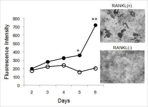
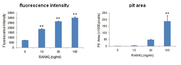
RANKL-dependent increase of fluorescence intensity of the conditioned medium (left) and pit area (right) from RAW264 cells in fluoresceinamine-labeled chondroitin sulfate (FACS)/ calcium phosphate (CaP)-coated plates (mean ± SD, n = 3, **: p<0.001).

骨吸收检测试剂盒                              Bone Resorption Assay Kit

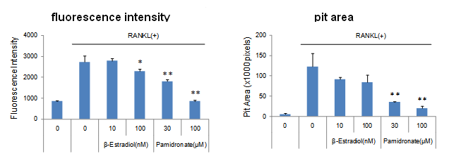
The inhibitory effects of Pamidronate and β-Estradiol on calcium phosphate resorption induced by RANKL (100 ng/mL) were measured by fluorescence intensity (left) and pit area (right) (mean ± SD, n = 3, *:p<0.05, **: p<0.001).

(摘自参考文献[4])

[1]

Chih-Hsin Kuo et al., arcinogenesis, 34. 11 2600-2609 (2013

[2]

Claire Fabre, Naoya Mimura, Katharyn Bobb, et al., Clin Cancer Res. 18, 4669-4681 (2012)

[3]

Kim J. et al., Cell. Biochem. 113:247-259 (2012)

[4]

Tatsuya Miyazaki. et al., Analytical Biochemistry 410 7-12 (2011)

[5]

Tatsuya Miyazaki. et al., Dental Materials Journal 29 4:403-410 (2010)

产品编号 产品名称 产品规格 产品等级
CSR-BRA-24KIT Bone Resorption Assay KIT 24 1 kit
CSR-BRA-48KIT Bone Resorption Assay KIT 48 1 kit
CSR-BRA-48X2KIT Bone Resorption Assay 48×2 2×1 KIT
CSR-BRA-24P Bone Resorption Assay Plate 24 1 PLATE
CSR-BRA-48P Bone Resorption Assay Plate 48 1 PLATE
CSR-BRA-48X2P Bone Resorption Assay Plate 48×2 2×1 PLATE
CSR-BRA-FACS1 Bone Resorption Assay FACS 13 mL
CSR-BRA-B1 Bone Resorption Assay Buffer 10 mL

LabAssay™ Ammonia LabAssay™ 氨检测试剂盒

  • 产品特性
  • 相关资料
  • Q&A
  • 参考文献

LabAssay™ Ammonia                              LabAssay™ 氨检测试剂盒

LabAssay Ammonia

LabAssay™ 氨检测试剂盒

氨主要产生于肠道和肾脏。在生物体内产生的氨通过肝脏中的尿素循环通路转变成尿素,然后从尿液中排出。

本产品把亚硝基铁氰化钠作为催化剂,以基于苯酚、次氯酸的贝特洛反应的比色法为基础的氨检测试剂。

LabAssay™ Ammonia 是以人和小鼠等动物样品作为对象的生化学检测用试剂,可检测样品中的氨。

LabAssay™ 系列是研究用试剂。不可用于诊断。

检测样品:血液

检测物种:小鼠、大鼠、人


◆特点


● 少量样品即可进行检测

● 可使用微孔板检测多个样品

● 可检测小鼠等动物样品


检测原理


  氨和苯酚在催化剂亚硝基铁氰化钠的作用下置换生成二氧二苯胺。在强碱存在下,利用与次氯酸钠反应生成呈蓝色的靛酚,通过测定吸光度,从而测定出样本中的氮氨浓度。


LabAssay™ Ammonia                              LabAssay™ 氨检测试剂盒


◆试剂盒组成


● 脱蛋白试剂——100 mL×1瓶

● 显色剂 A——50 mL×1瓶

● 显色剂 B——25 mL×1瓶

● 显色剂 C——50 mL×1瓶

● 氨标准溶液——15 mL×1瓶

● 标准溶液用稀释液——200 mL×1瓶


性能


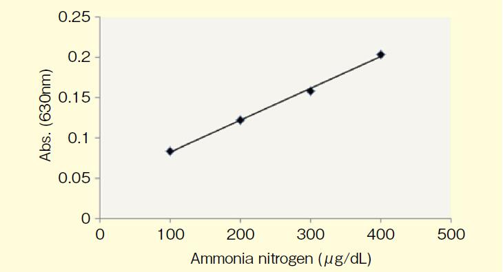
● 标准曲线范围——100~400 μg/dL(μg/100 mL)

● 检测时间——约70分钟

● 所需样品量——70 μL

● 检测波长——630 nm


〈标准曲线示例〉


LabAssay™ Ammonia                              LabAssay™ 氨检测试剂盒


◆使用方法


【微孔板法】

添加经脱蛋白试剂处理的样品 70 μL、显色剂 A 70 μL、显色剂 B 35 μL、显色剂 C 70 μL 至微孔板,使用孔板混匀仪充分混合,在37°C下加热 20 min。再次充分混合,室温下静置 30 min。以空白为对照,检测样品和标准溶液的吸光度。


【试管法】

添加经脱蛋白试剂处理的样品 800 μL、显色剂 A 800 μL、显色剂 B 400 μL、显色剂 C 800 μL 至微管或试管,充分搅拌,并在37°C下加热 20 min。再次搅拌后,用水(20~25°C)冷却 30 min。以空白为对照,检测样品和标准溶液的吸光度。

    使用此方法在试管内检测时,试剂盒的使用次数为 60 次。

◆产品列表


产品编号

产品名称

规格

包装

633-51761

(旧编号:295-78901)

LabAssay™ Ammonia

LabAssay™ 氨检测试剂盒

细胞生物学用

700 tests


相关产品


产品编号

产品名称

规格

包装

633-51021

(旧编号:291-58601)

LabAssay™ ALP

LabAssay™ 碱性磷酸酶检测试剂盒

细胞生物学用

500 tests

635-50981

(旧编号:294-65801

LabAssay™ Cholestrol

LabAssay™ 胆固醇检测试剂盒

细胞生物学用

500 tests

636-51011

(旧编号:290-65901)

LabAssay™ Creatinine

LabAssay™ 肌酸检测试剂盒

细胞生物学用

500 tests

638-50971

(旧编号:298-65701)

LabAssay™ Glucose

LabAssay™ 葡萄糖检测试剂盒

细胞生物学用

500 tests

633-52001

(旧编号:294-63601)

LabAssay™ NEFA

LabAssay™ 游离脂肪酸检测试剂盒

细胞生物学用

500 tests

639-51001

(旧编号:296-63801)

LabAssay™ Phospholipid

LabAssay™ 磷脂检测试剂盒

细胞生物学用

500 tests

632-50991

(旧编号:290-63701)

LabAssay™ Triglyceride

LabAssay™ 甘油三酯检测试剂盒

细胞生物学用

350 tests


点击了解LabAssay™全系列

产品编号 产品名称 产品规格 产品等级

非临床研究用检测试剂 免疫·过敏研究领域

  • 产品特性
  • 相关资料
  • Q&A
  • 参考文献

非临床研究用检测试剂                              免疫·过敏研究领域


免疫球蛋白E(IgE)

生产商编号
和光编号
产品名称 检测范围

检测样本量

(稀释样本)

AKRIE-010

633-02899

LBIS® IgE Mouse ELISA Kit  
小鼠免疫球蛋白E(IgE) ELISA试剂盒 
1~100 ng/mL 50 μL
AKRIE-011
636-04349

LBIS® IgE Rat ELISA Kit  
大鼠免疫球蛋白E(IgE)ELISA试剂盒

1~100 ng/mL 50 μL


◆白细胞介素(Interleukin)

生产商编号

和光编号

产品名称 检测范围 检测样本量
(稀释样本)
AKMIL12-011
638-40841
LBIS® Mouse IL-12 ELISA   Kit
小鼠白细胞介素-12(IL-12)ELISA试剂盒
2.87~700 pg/mL 100 μL

TDAR(T cell Dependent Antibody Responce)试验用


生产商编号
和光编号
产品名称 检测范围 检测样本量
(稀释样本)
AKRKG-010
632-13751

LBIS® KLH(TDAR)-Rat IgG ELISA KIT
血蓝蛋白(KLH)(T细胞依赖性抗原)

大鼠免疫球蛋白G(IgG) ELISA试剂盒 (*1)

0.47~30 ng/mL 50 μL
AKRKM-010
639-13761

LBIS® KLH(TDAR)-Rat IgM ELISA KIT
血蓝蛋白(KLH)(T细胞依赖性抗原)

大鼠免疫球蛋白M(IgM) ELISA试剂盒 (*2)

3.13~200 ng/mL 50 μL


(*1) 为了避免非特异性反应,请将检测样本稀释500倍以上

(*2) 为了避免非特异性反应,请将检测样本稀释200倍以上


抗OVA(ovalbumin)抗体

生产商编号
和光编号
产品名称 检测范围 检测样本量
(稀释样本)
AKRIE-030
639-07651

LBIS® OVA-IgE Mouse
小鼠卵清蛋白特异性免疫球蛋白E

OVA-IgE)ELISA试剂盒 

1.88~120 U/mL 10 μL
AKRIE-040
636-07661

LBIS® OVA-IgG1 Mouse
小鼠卵清蛋白特异性免疫球蛋白G1

OVA-IgG1)ELISA试剂盒

1.88~120 U/mL 10 μL


◆抗核酸抗体


生产商编号
和光编号
产品名称 检测范围 检测样本量
(稀释样本)
AKRDD-061
637-02691
LBIS® Anti dsDNA-Mouse ELISA Kit
小鼠抗双链DNA抗体 ELISA试剂盒
15.6~1000mU/mL 100 μL

AKRSD-051
630-02701

LBIS® Anti ssDNA-Mouse ELISA Kit
LBIS® 抗ssDNA-小鼠ELISA试剂盒 
15.6~1000mU/mL 100 μL

类风湿因子


生产商编号
和光编号
产品名称 检测范围 检测样本量
(稀释样本)
AKRRG-101
637-02679

LBIS® Rheumatoid Factor IgG-Mouse ELISA Kit
小鼠免疫球蛋白G(IgG)

类风湿因子 ELISA试剂盒

15.6~1000 mU/mL 100 μL
AKRRG-111
634-02689

LBIS® Rheumatoid Factor IgM-Mouse ELISA Kit
小鼠免疫球蛋白M(IgM)

类风湿因子 ELISA试剂盒

15.6~1000 mU/mL 100 μL

参考文献

◆LBIS® 小鼠免疫球蛋白E(IgE) ELISA试剂盒相关参考文献


 1.

Inhibitory Effect of Carnosol on Phthalic Anhydride-Induced Atopic Dermatitis via Inhibition of STAT3. Lee DY, Hwang CJ, Choi JY, Park MH, Song MJ, Oh KW, Son DJ, Lee SH, Han SB, Hong JT. Biomol Ther (Seoul). 2017 Sep 1;25(5):535-544.


 2.

Effects of aqueous extract from red Liriope platyphylla on phthalic-anhydride-induced atopic dermatitis in Interleukin-4/Luciferase/Consensus non-coding sequence-1 transgenic mice evaluated in terms of luciferase signal and general phenotype biomarkers. Kwak MH, Kim JE, Go J, Son HJ, Lee HS, Hong JT, Hwang DY. Journal of Traditional Chinese Medicine, Vol.37, Issue 4, Aug 2017, p475-485


 3.

Oral administration of freeze-dried powders of honey bee larvae inhibits the development of atopic dermatitis-like skin lesions in NC/Nga mice. Kageyama M, Xing G, Li K, Zhang Z, Sugiyama A. Personalized Medicine Universe, Vol. 6, July 2017, Pages 22-27.


 4.

Differential Nitric Oxide-Derived Protein Modifications of Flavin-Containing Monooxygenases in Type 1 Allergic Mice. Tanino T, Bando T, Komada A, Nojiri Y, Okada Y, Ueda Y, and Sakurai E. Drug Metabolism and Disposition  July 31, 2017,  dmd.117.076570;


 5.

Simultaneously increased expression of glucocorticoid‑induced tumor necrosis factor receptor and its ligand contributes to increased interleukin‑5/13‑producing group 2 innate lymphocytes in murine asthma. Zhang M, Wan J, Xu Y, Zhang D, Peng J, Qi C, Guo Q, Xia S, Su Z, Wang S, Xu H. Mol Med Rep. 2017 Jun;15(6):4291-4299.


 6.

Transgenic rat model of childhood-onset dermatitis by overexpressing telomerase reverse transcriptase (TERT). Kaneko R, Sato A, Hamada S, Yagi T, Ohsawa I, Ohtsuki M, Kobayashi E, Hirabayashi M, Murakami T. Transgenic Res. 2016 Aug;25(4):413-24.


 7.

Effects of sublingual immunotherapy in a murine asthma model sensitized by intranasal administration of house dust mite extracts. Shima K, Koya T, Tsukioka K, Sakagami T, Hasegawa T, Fukano C, Ohashi-Doi K, Watanabe S, Suzuki E, Kikuchi T. Allergol Int. 2016 Jul 8.


 8.

Therapeutic effect of tyndallized Lactobacillus rhamnosus IDCC 3201 on atopic dermatitis mediated by down-regulation of immunoglobulin E in NC/Nga mice. Lee SH, Yoon JM, Kim YH, Jeong DG, Park S, Kang DJ. Microbiol Immunol. 2016 Jul;60(7):468-76.


 9.

Gain-switched 311-nm Ti:Sapphire laser might be a potential treatment modality for atopic dermatitis.. Choi SY, Oh CT, Kwon TR, Kwon HJ, Choi EJ, Jang YJ, Kim HS, Chu H, Mun SK, Kim MN, Kim BJ. Lasers Med Sci. 2016 Jul 9.


10.

Perilla leaf extract prevents atopic dermatitis induced by an extract of Dermatophagoides farinae in NC/Nga mice. Komatsu KI, Takanari J, Maeda T, Kitadate K, Sato T, Mihara Y, Uehara K, Wakame K. Asian Pac J Allergy Immunol. 2016 Mar 20.


11.

Functional polysaccharides from Grifola frondosa aqueous extract inhibit atopic dermatitis-like skin lesions in NC/Nga mice. Park HS, Hwang YH, Kim MK, Hong GE, Lee HJ, Nagappan A, Yumnam S, Kim EH, Heo JD, Lee SJ, Won CK, Kim GS. Biosci Biotechnol Biochem. Vol.79(1), p147-54, 2015.


12.

Inhibitory effect of 5,6-dihydroergosteol-glucoside on atopic dermatitis-like skin lesions via suppression of NF-κB and STAT activation. Jung M, Lee TH, Oh HJ, Kim H, Son Y, Lee EH, Kim J. J Dermatol Sci. 2015


13.

Immunopotentiator from Pantoea agglomerans Prevents Atopic Dermatitis Induced by Dermatophagoides farinae Extract in NC/Nga Mouse. Wakame K, Komatsu K, Inagawa H, Nishizawa T. Anticancer Res. Vol.35(8), p4501-8, Aug 2015.


14.

Solanum tuberosum L. cv Jayoung Epidermis Extract Inhibits Mite Antigen-Induced Atopic Dermatitis in NC/Nga Mice by Regulating the Th1/Th2 Balance and Expression of Filaggrin. Yang G, Cheon SY, Chung KS, Lee SJ, Hong CH, Lee KT, Jang DS, Jeong JC, Kwon OK, Nam JH, An HJ. J Med Food. 2015 Jun 23.


15.

Gamisasangja-tang suppresses pruritus and atopic skin inflammation in the NC/Nga murine model of atopic dermatitis. Park BK, Park YC, Jung IC, Kim SH, Choi JJ, Do M, Kim SY, Jin M. J Ethnopharmacol. 2015 Feb 23.


16.

Bacillus subtilis KCTC 11782BP-produced alginate oligosaccharide effectively suppresses asthma via T-helper cell type 2-related cytokines. Bang MA, Seo JH, Seo JW, Jo GH, Jung SK, Yu R, Park DH, Park SJ. PLoS One. 2015 Feb 6;10(2):e0117524.


17.

Effect of the hand antiseptic agents benzalkonium chloride, povidone-iodine, ethanol, and chlorhexidine gluconate on atopic dermatitis in NC/Nga mice. Sadakane K, Ichinose T. Int J Med Sci. Vol.5;12(2), p116-25, Jan 2015.


18.

Effects of oral administration of di-(2-ethylhexyl) and diisononyl phthalates on atopic dermatitis in NC/Nga mice. Sadakane K., Ichinose T., Takano H., Yanagisawa R. and Koike E. Immunopharmacology and Immunotoxicology, Vol.36(1), p61-69, Feb 2014.


19.

Oral Administration of SSC201, a Medicinal Herbal Formula, Suppresses Atopic Dermatitis-Like Skin Lesions. Kyung P., Chun P., Chul J., Hyung K., Eun C., Sunyoung P., June C. and Mirim J. Journal of Medicinal Food, 2014.


20.

Effect of Allergen Sensitization on External Root Resorption. Murata N., Ioi H., Ouchi M., Takao T., Oida H., Aijima R., Yamaza T., Kido MA. JDR, Vol.92(7), p641-647, Jul 2013.


21.

Quantitative evaluation of therapeutic effect of Liriope platyphylla on phthalic anhydride-induced atopic dermatitis in IL-4/Luc/CNS-1 Tg mice. M.H.Kwak, J.E.Kim, I.S.Hwang, Y.J.Lee, B.S.An, J.T.Hong, S.H.Lee, D.Y.Hwang. Journal of Ethnopharmacology, Vol.148(3), p880-889, Jul 2013.

22.

Lack of both α2-antiplasmin and plasminogen activator inhibitor type-1 induces high IgE production. K.Okada, S.Ueshima, N.Kawao, M.Yano, Y.Tamura, M.Tanaka, A.Sakamoto, M.Hatano, M.Arima, S.Miyata, N.Nagai, T.Tokuhisa, O.Matsuo. Life Sciences, Vol.93(2-3), p89-95, Jul 2013.

23.

Therapeutic effects of fermented soycrud on phenotypes of atopic dermatitis induced by phthalic anhydride. Sung J-E., Kwak M-H., Kim J-E., Lee Y-J., Kim R-U., Kim E-A., Lee G-Y., Kim D-S. and Hwang D-Y. Lab Anim Res, Vol.29(2), p103-112, Jun 2013.

24.

Water-soluble chitosan and herbal honey compound alleviates atopic dermatitis-like lesions in NC/Nga mice. C.Choi, W.-S.Kim, Y.H.Park, S.-C.Park, M.-K.Jang, J.-W.Nah. Journal of Industrial and Engineering Chemistry, May 2013.


25.

Interleukin-17/T-helper 17 cells in an atopic dermatitis mouse model aggravate orthodontic root resorption in dental pulp. M.Shimizu, M.Yamaguchi, S.Fujita, T.Utsunomiya, H.Yamamoto, K.Kasai. European Journal of Oral Sciences, Vol.121(2), p101-110, Apr 2013.


26.

Water-soluble chitosan and herbal honey compound alleviates atopic dermatitis-like lesions in NC/Nga mice. Choi C., Kim W-S., Park YH., Park S-C., Jang M-K., Nah J-W. Journal of Industrial and Engineering Chemistry, Vol.20(2), p499-504, Mar 2014.


27.

Indirubin, a purple 3,2- bisindole, inhibited allergic contact dermatitis via regulating T helper (Th)-mediated immune system in DNCB-induced model. Kim M H, Choi Y Y, Yang G, Cho I-H, Nam D, Yang W M. Journal of Ethnopharmacology, Vol.145(1), p214-219, Jan 2013.


28.

Effects of Catalpa ovata stem bark on atopic dermatitis-like skin lesions in NC/Nga mice. Yang G, Choi C-H, Lee K, Lee M, Ham I, Choi H-Y. Journal of Ethnopharmacology, Vol.145(2), p416-423, Jan 2013.


29.

Interleukin 33 Is Induced by Tumor Necrosis Factor α and Interferon γ in Keratinocytes and Contributes to Allergic Contact Dermatitis. Taniguchi K., Yamamoto S., Hitomi E., Inada Y., Suyama Y., Sugioka T., Hamasaki Y. J Investig Allergol Clin Immunol, Vol.23(6), p428-434, 2013.


30.

Organic Chemicals in Diesel Exhaust Particles Enhance Picryl Chloride-Induced Atopic Dermatitis in NC/Nga Mice. Sadakane K., Ichinose T., Takano H., Yanagisawa R., Inoue K., Kawazato H., Yasuda A., Hayakawa K. Int Arch Allergy Immunol, Vol.162(1), p7-15, 2013.


31.

The effects of (±)-Praeruptorin A on airway inflammation, remodeling and transforming growth factor-β1/Smad signaling pathway in a murine model of allergic asthma. Xiong Y, Wang J, Wu F, Li J, Kong L. International Immunopharmacology, Vol.14(4), p392-400, Dec 2012.


32.

Inhibitory effects of Drynaria fortunei extract on house dust mite antigen-induced atopic dermatitis in NC/Nga mice. Sung Y-Y, Kim D-S, Yang W-K, Nho K J, Seo H S, Kim Y S, Kim H K.. Journal of Ethnopharmacology, Vol.144(1), p94-100, Oct 2012.


33.

Topical application of an ethanol extract prepared from Illicium verum suppresses atopic dermatitis in NC/Nga mice. Sung Y-Y, Yang W-K, Lee A Y, Kim D-S, Nho K J, Kim Y S, Kim H K.. Journal of Ethnopharmacology, Vol.144(1), p151-159, Oct 2012.


34.

Anti-allergic effect of lactic acid bacteria isolated from seed mash used for brewing sake is not dependent on the total IgE levels. Masuda Y, Takahashi T, Yoshida K, Nishitani Y, Mizuno M, Mizoguchi H. Journal of Bioscience and Bioengineering, Vol.114(3), p292-296, Sep 2012.


35.

Advantages of histamine H4 receptor antagonist usage with H1 receptor antagonist for the treatment of murine allergic contact dermatitis. Matsushita A, Seike M, Okawa H, Kadawaki Y, Ohtsu H. Experimental Dermatology, Vol.21(9), p714-715, Sep 2012.


36.

Platycodon grandiflorus alleviates DNCB-induced atopy-like dermatitis in NC/Nga mice. Park S-J, Lee H-A, Kim J W, Lee B-S and Kim E-J. Indian J Pharmacol. Vol.44(4), p469-474, Jul-Aug 2012.


37.

Effect of Chrysanthemi borealis flos on atopic dermatitis induced by 1-chloro 2,4-dinitrobenzene in NC/Nga mouse. Yang G, Lee K, An D-G, Lee M-H, Ham I-H, Choi H-Y. Immunopharmacology and Immunotoxicology, Vol.34(3), p413-418, Jun 2012.


38.

Effects of (±)-praeruptorin A on airway inflammation, airway hyperresponsiveness and NF-κB signaling pathway in a mouse model of allergic airway disease. Xiong Y, Wang J, Wu F, Li J, Zhou L, Kong L. European Journal of Pharmacology, Vol.683(1-3), p316-324, May 2012.


39.

Anti-allergic effect of lactic acid bacteria isolated from seed mash used for brewing sake is not dependent on the total IgE levels. Y. Masuda., T. Takahashi., K. Yoshida., Y. Nishitani., M. Mizuno., H. Mizoguchi. Journal of Bioscience and Bioengineering, 2012.


40.

The Protective and Recovery Effects of Fish Oil Supplementation on Cedar Pollen-Induced Allergic Reactions in Mice. Hirao A, Furutani N, Nagahama H, Itokawa M, Shibata S. Food and Nutrition Sciences, Vol.3, p40-47, 2012.


41.

Th17-cells in atopic dermatitis stimulate orthodontic root resorption. Yamada K, Yamaguchi M, Asano M, Fujita S, Kobayashi R, Kasai K. Oral Diseases, 2012.


42.

Mixture of Polyphenols and Anthocyanins from Vaccinium uliginosum L. Alleviates DNCB-Induced Atopic Dermatitis in NC/Nga Mice. Kim M J and Choung S-Y. Evidence-Based Complementary and Alternative Medicine, Vol.2012 (2012)


43.

Single systemic administration of Ag85B of mycobacteria DNA inhibits allergic airway inflammation in a mouse model of asthma. Karamatsu K, Matsuo K, Inada H, Tsujimura Y, Shiogama Y, Matsubara A, Kawano M and Yasutomi Y. J Asthma Allergy, Vol.5, p71-79, 2012.


44.

Inhibitory Effect of Nelumbo nucifera (Gaertn.) on the Development of Atopic Dermatitis-Like Skin Lesions in NC/Nga Mice. R. Karki., M.-A Jung., K.-J. Kim., and D.-W Kim. Evidence-Based Complementary and Alternative Medicine, Vol. 2012 (2012), Article ID 153568, 7 pages


45.

Lack of Transient Receptor Potential Vanilloid-1 Enhances Th2-Biased Immune Response of the Airways in Mice Receiving Intranasal, but Not Intraperitoneal, Sensitization. T. Mori., K. Saito., Y. Ohki., H. Arakawa., M. Tominaga., K. Tokuyama. Int Arch Allergy Immunol 2011, Vol.156, p305-312.


46.

Improvement of the Antiinflammatory and Antiallergic Activity of Bidens pilosa L. var. radiata SCHERFF Treated with Enzyme (Cellulosine). Horiuchi, M. and Seyama, Y. Journal of Health Science, Vol. 54(2008), No.3, p294-301.


47.

Suppression of spontaneous dermatitis in NC/Nga murine model by PG102 isolated from Actinidia arguta. Park, E.J., Park, K.C., Eo, H, Seo, J., Son, M., Kim, K.H. et al. J Investigative Dermatology, Vol.127, p1154-1160, 2007.


48.

Sopungyangjae-Tang inhibits development of dermatitis in Nc/Nga mice. Pokharel, Y.R., Lim, S.C., Kim, S.C., Heo, T.H., Choi, H.K. and Kang, K.W. Evidence-based Complementary and Alternative Medicine nem015, 1-8, March 12, 2007.


49.

Inhibitory effect of DA-9201, an extract of Oryza sativa L., on airway inflammation and bronchial hyperresponsiveness in mouse asthma model. Lee, S.H., Sohm, Y.S., Kang, K.K., Kwon, J.W. and Yoo, M. Biol. Pharm. Bull. Vol.29(6), p1148-1153, 2006.


50.

A new symbiotic, Lactobacillus casei subsp. casei together with dextran, reduces murine and human allergic reaction. Ogawa, T., Hashikawa, S., Asai, Y., Sakamoto, H., Yasuda, K., and Makimura, Y. FEMS Immunology and Medical Microbiology, Vol.46, p400-409, 2006.


51.

Effects of alpha tocopherol and probucol supplements on allergen-induced airway inflammation and hyperresponsiveness in a mouse model of allergic asthma. Okamoto, N., Murata, T., Tamai, H., Tanaka, H., and Nagai, H. Int. Allergy Immunol. Vol.141, p172-180, 2006.


52.

Control of IgE and selective TH1 and TH2 cytokines by PG102 isolated from Actinidia arguta. Parkm E.J., Kim, B., Eo, H., Park, K, Kim, Y., Lee, H.J. and Son, M. J Allergy and Clinical Immunol, p1151-1167, 2005.


53.

Oral immunotherapy against a pollen allergy using a seed-based peptide vaccine. Takagi, H., Saito, S., Yang, L., Nagasawa, S., Nishizawa, N., and Takaiwa, F. Plant Biotechnology Journal, Vol.3, p521-533, 2005.


54.

Activated protein C inhibits bronchial hyperresponsiveness and Th2 cytokine expression in mice. Hisamichi Yuda, Yukihiko Adachi, Osamu Taguchi, Esteban C. Gabazza, Osamu Hataji, Hajime Fujimoto, Shigenori Tamaki, Kimiaki Nishikubo, Kenji Fukudome, Corina N. D’Alessandro-Gabazza, Junko Maruyama, Masahiko Izumizaki, Michiko Iwase, Ikuo Homma, Ryo Inoue, Haruhiko Kamada, Tatsuya Hayashi, Michael Kasper, Bart N. Lambrecht, Peter J. Barnes, and Koji Suzuki. Blood, Vol.103, No.6, p2196-2204, 2004.


55.

Inadequate induction of suppressor of cytokine signaling-1 causes systemic autoimmune diseases. Fujimoto, M., Tsutsui, H., Xinshou, O., Tokumoto, M., Watanabe, D., Shima, Y., Yoshimoto, T., Hirakata, H., Kawase, I., Nakanishi, K., Kishimoto, T., and Naka, T. International Immunology Vol.16, p303-314, 2004.


56.

Association of cathepsin E deficiency with development of atopic dermatitis. Tsukuba, T., Okamoto, K., Okamoto, Y., Yanagawa, M. J Biochemistry, Vol.134, p893-902, 2003.


57.

Mast-cell-dependent histamine release after praziquantel treatment of Schistosoma japonicum infection: implications for chemotherapy-related adverse effects. Jun Matsumoto and Hajime Matsuda. Parasitology Research, Vol.88, p888 – 893, 2002.


◆LBIS® 大鼠免疫球蛋白E(IgE) ELISA试剂盒相关参考文献


1.

Transgenic rat model of childhood-onset dermatitis by overexpressing telomerase reverse transcriptase (TERT). Ryosuke Kaneko, Atsuko Sato, Shun Hamada, Takeshi Yagi, Ichiro Ohsawa, Mamitaro Ohtsuki, Eiji Kobayashi, Masumi Hirabayashi, Takashi Murakami. Transgenic Research, pp 1-12, First online: 17 February 2016


2.

Oral administration of drugs with hypersensitivity potential induces germinal center hyperplasia in secondary lymphoid organ/tissue in Brown Norway rats, and this histological lesion is a promising candidate as a predictive biomarker for drug hypersensitivity occurrence in humans. A.Tamura, I.Miyawaki, T.Yamada, J.Kimura, H.Funabashi. Toxicology and Applied Pharmacology, Vol.271(1), p30-40, Aug 2013.


3.

Enhancement of the pulmonary allergic granulocyte recruitment in rats exposed to DMTI-Ⅱ,a Kunitz-type inhibitor isolated from Dimorphandra mollis seeds. Glaucia,C,M.,Karina,F,R.,Dalize,M,S.,Andre,A,S.,Ivani,A,de S.,Maria,L R,M.,Edson,A.. International Immunopharmacology, Vol.11(6),  p740-747, Jun 2011.


4.

Altered cytokine expression in mesenteric lymph nodes in a rat strain (Matsumoto Eosinophilic Shinshyu) that spontaneously develops hypereosinophilia. Muto, S., Monnai, M., Okuhara, Y., Murakami, M., Kuroda, J., Ono, T., and Matsumoto, K.. Immunology, Vol.116, p373-380, 2005.


◆LBIS® 小鼠免疫球蛋白G(IgG)类风湿因子ELISA试剂盒相关参考文献


 1.

Dysbiosis Contributes to Arthritis Development via Activation of Autoreactive T Cells in the Intestine. Maeda Y, Kurakawa T, Umemoto E, Motooka D, Ito Y, Gotoh K, Hirota K, Matsushita M, Furuta Y, Narazaki M, Sakaguchi N, Kayama H, Nakamura S, Iida T, Saeki Y, Kumanogoh A, Sakaguchi S, Takeda K. Arthritis Rheumatol. 2016 Nov;68(11):2646-2661.


 2.

C9orf72 ablation causes immune dysregulation characterized by leukocyte expansion, autoantibody production, and glomerulonephropathy in mice. Atanasio A, Decman V, White D, Ramos M, Ikiz B, Lee HC, Siao CJ, Brydges S, LaRosa E, Bai Y, Fury W, Burfeind P, Zamfirova R, Warshaw G, Orengo J, Oyejide A, Fralish M, Auerbach W, Poueymirou W, Freudenberg J, Gong G, Zambrowicz B, Valenzuela D, Yancopoulos G, Murphy A, Thurston G, Lai KM. Sci Rep. 2016 Mar 16


 3.

Reciprocal effects of Schistosoma mansoni infection on spontaneous autoimmune arthritis in IL-1 receptor antagonist-deficient mice. Osada Y, Yamada S, Nakae S, Sudo K, Kanazawa T. Parasitol Int. Vol.64(1), p13-17. Feb 2015.


 4.

The development of mixed cryoglobulinemia in Capillaria hepatica-infected mice is associated with the capillaria antigen-induced selective proliferation of splenic B-1a cells in response to interleukin-5 stimulation. Aihara N, Kamiie J, Yamada M, Shirota K. Am J Pathol. Vol.185(1), p172-84, Jan 2015.


 5.

Mechanism of the 24-hour rhythm of tumor necrosis factor-alpha formed by onset of rheumatoid arthritis. Yoshimatsu H., Okazaki F., Ieiri I., To H. Chronobiology International, 2014.


 6.

Deficient Leptin Signaling Ameliorates Systemic Lupus Erythematosus Lesions in MRL/Mp-Faslpr Mice. Fujita Y., Fujii T., Mimori T., Sato T., Nakamura T., Iwao H., Nakajima A., Miki M., Sakai T., Kawanami T., Tanaka M., Masaki Y., Fukushima T., Okazaki T. and Umehara H. The Journal of Immunology, Vol.192(3), p979-984, Feb 2014.


 7.

Phenotype conversion from rheumatoid arthritis to systemic lupus erythematosus by introduction of Yaa mutation into FcγRIIB-deficient C57BL/6 mice. Kawano S, Lin Q, Amano H, Kaneko T, Nishikawa K, Tsurui H, Tada N, Nishimura H, Takai T, Shirai T, Takasaki Y, Hirose S. European Journal of Immunology, Vol.43(3), p770-778, Mar 2013.


 8.

IFN-γ-Producing Effector CD8 T Lymphocytes Cause Immune Glomerular Injury by Recognizing Antigen Presented as Immune Complex on Target Tissue. Tsumiyama K., Hashiramoto A., Takimoto M., Tsuji-Kawahara S., Miyazawa M. and Shiozawa S. The Journal of ImmunologyJuly, Vol.191(1), p91-96, 2013.


 9.

Increased expression of soluble form of vascular cell adhesion molecule-1 aggravates autoimmune arthritis in MRL-Fasipr mice. Oishi, H., Mizuki, S., Terada, M., Kudo, M., Araki, K., Araki, M., Nose, M., and Takahashi, S. Pathology International, Vol.57, p734-740, 2007.


10.

Electro-acupuncture at acupoint ST36 reduces inflammation and regulates immune activity in collagen-induced arthritic mice. Yim, Y.Y., Lee, H., Hong, K.E., Kim, Y.I., Lee, B.R., Son, C.G., and Kim, J.E. Evidence-based Complementary and Alternative Medicine 4:p51-57, 2007.

11.

Requirement for CD100-CD72 interactions in fine-tuning of B-cell antigen receptor signaling and homeostatic maintenance of the B-cell compartment. Kumanogoh, A., Shikina, T., Watanabe, C., Takegahara, N., Suzuki, K., Yamamoto, M., Takamatsu, H., Prasad, D.V.R., Mizui, M., Toyohuku, T., Tamaru, M., Watanabe, D., Parnes, J.R., and Kikutani, H. International Immuno, Vol.17, p1277-1282, 2005.


12.

The point mutation of tyrosine 759 of the IL-6 family cytokine receptor gp130 synergizes with HTLV-1 pX in promoting rheumatoid arthritis-like arthritis. Ishihara, K., Sawa, S., Ikushima, H., Hirota, S., Atsumi, T., Kamimura, D., Park, S.J., Murakami, M., Kitanuma, Y., Iwakura, Y., and Hirano, T. International Immunol. Vol.16, p455-465, 2004.


13.

Chemical induction of HO-1 suppresses lupus nephritis by reducing local iNOS expression and synthesis of anti-dsDNA antibody. Takeda, Y., Takeno, M., Iwasaki, M., Kobayashi, H., Kirino, Y., Ueda, A., Nagahama, K., Aoki, I., and Ishigatsubo, Y. Clin Exp Immunol. Vol.138, p237-244, 2004.


14.

A Point Mutation of Tyr-759 in Interleukin 6 Family Cytokine Receptor Subunit gp130 Causes Autoimmune Arthritis. T. Atsumi., K. Ishihara., D. Kamimura., H. Ikushima., T. Ohtani., S. Hirota., H. Kobayashi., S. Park., Y. Saeki., Y. Kitamura. and T. Hirano. J Exp Med, 196, Number 7, p979-990, 2002.


15.

A novel assay kits for autoantibodies rate on spontaneous autoimmune model mice. Kikukawa, T, Kojima, M., and Abe, C. Jap J Inflammation, Vol.20, p697-701, 2000.


◆LBIS® 小鼠免疫球蛋白M(IgM)类风湿因子ELISA试剂盒相关参考文献


 1.

C9orf72 ablation causes immune dysregulation characterized by leukocyte expansion, autoantibody production, and glomerulonephropathy in mice.
Atanasio A, Decman V, White D, Ramos M, Ikiz B, Lee HC, Siao CJ, Brydges S, LaRosa E, Bai Y, Fury W, Burfeind P, Zamfirova R, Warshaw G, Orengo J, Oyejide A, Fralish M, Auerbach W, Poueymirou W, Freudenberg J, Gong G, Zambrowicz B, Valenzuela D, Yancopoulos G, Murphy A, Thurston G, Lai KM.

Sci Rep. 2016 Mar 16

 2.

Production of Autoantibodies by Murine B-1a Cells Stimulated with Helicobacter pylori Urease through Toll-Like Receptor 2 Signaling
Kobayashi F, Watanabe E, Nakagawa Y, Yamanishi S, Norose Y, Fukunaga Y and Takahashi H.

Infect. Immun, Vol.79(12), p4791-4801, Dec 2011.


 3.

Lack of B and T lymphocyte attenuator exacerbates autoimmune disorders and induces Fas-independent liver injury in MRL-lpr/lpr mice
Y. Oya., N. Watanabe. Y. Kobayashi., T. Owada., M. Oki., K. Ikeda. A. Suto., S. Kagami., K. Hirose., T. Kishimoto., and H. Nakajima.

Int. Immunol. Vol.23(5), p335-344, 2011.


 4.

An experimental type 2 mixed cryoglobulinemia with renal glomerulopathy in ICR mice triggered by Capillaria hepatica infection
N. Aihara., J. K. Chambers., K. Hosono., J. Kamiie., and K. Shirota.

Histol Histopathol, Vol.26, p1519-1529, 2011.


 5.

Point mutation of tyrosine 759 of the IL-6 family cytokine receptor,gp130,augments collagen-induced arthritis in DBA/IJ mice
Tsuji,F.,Yoshimi,M.,Katsuta,O.,Takai,M.,Ishihara,K.,Aono,H.

BMC Musculoskeletal Disorders ,10:23, 2009.


 6.

Implications for induction of autoimmunity via activation of B-1 cells by Helicobacter pylori urease.
Yamasnishi, S., Iizumi, T., Watanabe, E., Shimizu, M., Kamiya, S., Nagata, K., Kumagai, Y., Fukunaga, Y., and Takahashi, H.

Infection and Immunity, Vol.74, p248-256, 2006.


 7.

Cytokine production profile of splenocytes derived from zymosan A-treated SKG mice developing arthritis.
Kobayashi, K., Suda, T., Nan-ya, K., Sakaguchi, N., Sakaguchi, S., and Miki, I.

Inflammation Research, Vol.55, p335-341, 2006.


 8.

Chronic polyarthritis caused by mammalian DNA that escapes from degradation in macrophages.
Kawane, K., Ohtani, M., Miwa, K., Kizawa, T., Kanbara, Y., Yoshioka, Y., and Yoshikawa,H.

Nature, Vol.443, p998-1002, 2006.


 9.

Requirement for CD100-CD72 interactions in fine-tuning of B-cell antigen receptor signaling and homeostatic maintenance of the B-cell compartment.
Kumanogoh, A., Shikina, T., Watanabe, C., Takegahara, N., Suzuki, K., Yamamoto, M., Takamatsu, H., Prasad, D.V.R., Mizui, M., Toyohuku, T., Tamaru, M., Watanabe, D., Parnes, J.R., and Kikutani, H.

International Immuno, Vol.17, p1277-1282, 2005.


10.

A Point Mutation of Tyr-759 in Interleukin 6 Family Cytokine Receptor Subunit gp130 Causes Autoimmune Arthritis
T. Atsumi., K. Ishihara., D. Kamimura., H. Ikushima., T. Ohtani., S. Hirota., H. Kobayashi., S. Park., Y. Saeki., Y. Kitamura. and T. Hirano

J Exp Med, 196, Number 7, p979-990, 2002.


11.

A novel assay kits for autoantibodies rate on spontaneous autoimmune model mice.
Kikukawa, T, Kojima, M., and Abe, C.

Jap J Inflammation, Vol.20, p697-701, 2000.


产品编号 产品名称 产品规格 产品等级

催产素检测试剂盒 Oxytocin ELISA kit

  • 产品特性
  • 相关资料
  • Q&A
  • 参考文献

Oxytocin ELISA kit 催产素检测试剂盒催产素检测试剂盒                              Oxytocin ELISA kit 催产素检测试剂盒                              Oxytocin ELISA kit


◆原理


催产素是神经垂体表达的一种多肽,分泌在下丘脑的室旁核,并储存在垂体后叶。催产素由9个氨基酸构成,有一个[1-6]二硫键和一个半胱氨酸。曾被认为是只作用于女性生殖的激素,现今实验证明,催产素也能作用于神经递质,可能也涉及到神经精神障碍,社交/性行为和对雄性生殖生理有着重要的作用。催产素和相关神经垂体多肽、精氨酸 -加压素,能维持肾积水和钠的平衡。

Enzo 提供的催产素 ELISA 检测试剂盒用比色法,过夜后只需一个小时就能出结果。吸光度为 405 nm。不检测加压素,检测结果可信度高。灵敏度高,可检测 15 pg/mL 的催产素。试剂用不同颜色标记,在实验室中使用时,能减少失误。

该试剂盒灵敏度高,特异性强,可快速、低成本的检测催产素含量。


◆优点特色


● 特异性强,不会检测加压素,检测结果可信度高。

● 灵敏度高,可检测 15 pg/mL 的催产素。

● 成本低,和 LC/MS 方法 相比,检测速度更快,花费更低。

● 试剂用不同颜色标记和预包被检测板,使用更方便,减少误操作。

● 大量文献报道

● 可提供大包装

催产素检测试剂盒                              Oxytocin ELISA kit

 ◆产品规格


灵敏度

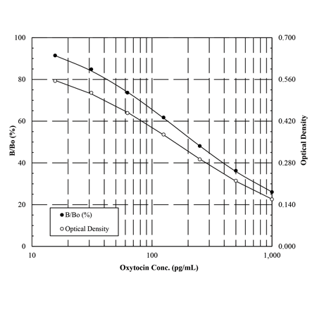
15 pg/mL (范围15.6 – 1,000 pg/mL)

检测时间

过夜后1小时

应用

ELISA,比色检测

样品

可检测培养上清液、牛奶、血浆、和所有物种的血清内催产素含量。

已验证的样品类型有:脑脊髓液、唾液、组织和尿液。

种属

无种属特异性

交叉反应

催产素(7%),催产素(100%),Arg8-催产素(7.5%),和其他相关分子<0.02%。

使用/稳定性

除了标准品和偶联物要存放在-20°C ,其余所有物质存放在4°C。

试剂盒的组成

GXR 微孔板,偶联物,抗体,分析缓冲液,洗涤缓冲液,标准品,pNpp 底物,终止液

UniProt   ID

P01178 (human)

催产素 ELISA 试剂盒 Q&A


Q:    催产素是否有必要抽提?

A:    Enzo Life Sciences 的催产素 ELISA 试剂盒需要从血清或者血浆样品中进行抽提。需要抽提是由于血清或者血浆样品中存在的基质干扰

          (matrix  reference),而不是因为预期的基础水平。其他样品没有证实过需要进行抽提。


Q:    如果血清或者血浆样品不进行抽提,是否还有另外的方法?

A:    以下方法是从液体中抽提催产素的实验流程:

    1.     添加2体积的预冷丙酮到样品中。震荡混匀。

    2.     离心 12,000 ×g,20 分钟。

    3.     将上清转移到新管中。

    4.     添加5倍体积的预冷石油醚。震荡混匀。

    5.     离心10,000×g,10 分钟。

    6.     弃上层的醚。小心转移留下的液体层到玻璃管,用氮气或者氩气进行干燥。

    7.     用 assay  buffer 重悬样品。


          请注意抽提处理过程中肽的回收率是变化的。需要对抽提步骤进行优化。确定抽提效率有很多种方法,包括放射性多肽,加样回收实验(添

          平行样品,检测已知浓度催产素的回收率)


Q:    是否可检测唾液样品?

A:    虽然在研发该试剂盒中没有检测过唾液样品,但是很多客户对检测唾液样品中的催产素感兴趣。有文献(点击此处)报道用该试剂盒检测

          唾液样品。有科研人员使用下列步骤检测唾液样品。请注意,需要对您检测的具体样品进行条件优化。


    1.     收集1mL 唾液。使用前样品需要保存在-20℃。

    2.     离心直到出现沉淀颗粒,使粘液粘附在管底。

    3.     收集上清,每1mL 进行分装。

    4.     用离心蒸发器(SpeedVac 或者类似)蒸发1mL。

    5.     加入 250 μL assay buffer 重悬干燥的样品。


          上述样品进行了4倍浓缩,应该不含有干扰因子。


Q:    我需要使用多少样品?建议的稀释比是多少?

A:    需要样品的量和建议的稀释比取决于样品中催产素的水平。建议客户参考相关的文献,最好是相近的样品类型,检测条件。


Q:    除了培养上清,牛奶,血浆或者血清外,还可以检测什么样品?

A:    文献报道有检测脑脊髓液、唾液、组织和尿液。


Q:    该试剂盒是否可以检测任意物种?

A:    虽然在一些灵长类动物中(点击此处)发现单一氨基酸替换的新型催产素(会影响抗体结合的亲和力),但是在大多数哺乳动物间催产素的

          氨基酸顺序没有发生大变化,因此该试剂盒可以检测任意物种。文献报道检测的物种包括狒狒,人,小鼠,猪,大鼠,猕猴,绢毛猴。请注

          意如果用兔血清检测会有问题。不是由于催产素本身,而是因为该试剂盒使用的是抗兔的 IgG,会引起高背景值。


Q:    使用什么柱子进行催产素的抽提?

A:    使用含有 200 mg 吸附剂、柱容量 3.5 mL 的反向 C18 柱进行固相抽提(SPE)。如果血清/血浆样品少的话,可以使用更小的 C18 柱。


Q:    应该如何处理,变成干燥的样品?

A:    使用真空离心(比如 Speed Vac)或者氮气或者氩气进行干燥样品。如果样品数量多,建议使用真空离心的方法。样品可室温放置在

          SpeedVac过夜。第二天样品会变干燥,保持了样品的完整度(在真空状态下没有与外界环境接触)。


          用氮气干燥的话,需要将玻璃枪头贴到气体的底部,将枪头正好悬在含有样品管子的正下方。如果使用这个方法,建议先用含有石油醚的管子

          进行练习,检测整个过程耗时多少。


          不建议将样品放在罩子下面进行干燥。样品会氧化,残留的溶剂会影响检测。


Q:    如何判断我的样品是需要进行稀释还是抽提?

A:    如果样品经过推荐的最小稀释比稀释后检测值在试剂盒的检测范围内(标准曲线的范围内),那就稀释后直接进行检测。如果样品低于检测的

          最小值,必须进行抽提和浓缩。


Q:    如何使用excel制作标准曲线?

A:    1.     计算标准品和样品的平均净 OD 值。
                  平均的净 OD 值=平均 OD- 平均 NSB OD(Average Net OD=Average OD- Average NSB OD)
          2.     计算每个标准品的结合(与最大结合孔 Bo 的百分比)
                  结合率=净 OD/净 Bo OD ×100(Percent Bound=Net OD/Net Bo OD ×100)
          3.     Excel 程序,y轴为结合率(percent bound),x轴是标准品的浓度。开始-插入-散点图-添加趋势
线-对数,显示R值和公式即可。

                  对数方程式。公式是 y = a*ln(x) + b,x = inv ln [(y – b) / (a)] 或者 x = e^[(y – b) / (a)] .

Q:    血液样品该如何收集?

A:    血液样品应该收集到冷冻血清或者含有抑肽酶(500 KIU/mL 血液)EDTA(1 mg/mL 血液)管。在4℃,1600×g 离心样品 15 分钟。

          将血清或者血浆转移到塑料管内,保存在-70℃ 或者更低的温度,避免反复冻融。


Q:    如果样品是血浆,是否可以先稀释样品后再检测?

A:    说明书上没有提供血浆样品稀释的最小稀释比,因此建议样品抽提后进行检测。


Q:    如果样品是血清,是否可以先稀释样品再进行检测?

A:    说明书建议人血清的最小稀释比是1:8。建议在1:2~1:16 范围之间摸索最佳的稀释比例(以稀释后测到的 OD 值落在标准曲线范围

          最佳)。如果上述实验结果不理想,建议对样品进行抽提。


Q:    如何评估样品抽提(样品前处理)的效果?

A:    制备加样样品(spiked sample),将 200 pg/mL 催产素(已知浓度的标准品)加到不同的样品类型中。然后样品和加样样品进行抽提、

          检测。同时制备标准曲线。抽提效率的公式如下:加样样品的浓度(参考标准曲线计算加样样品的浓度)—抽提后样品的浓度/200pg/mL

          (理论浓度) ×100%。当抽提效率在 80%~120% 时,抽提效果最佳。


【参考文献

[1]

A sexually dimorphic hypothalamic circuit controls maternal care and oxytocin secretion: N. Scott, et al.; Nature (2015), Application(s): ELISA using isoflurane and blood samples from mice


[2]

Conspecific recognition and aggression reduction to familiars in newly weaned, socially plastic mammals: K.J. Robinson, et al.; Behav. Ecol. Sociobiol. 59, 1383 (2015),Application(s): ELISA using seal pup plasma

[3]

Electro-acupuncture improves the social interaction behavior of rats: H.F. Zhang, et al.; Physiol. Behav. 151, 485 (2015), Application(s): ELISA using rat brain tissue

[4] 

Oxytocin increases VTA activation to infant and sexual stimuli in nulliparous and postpartum women: R. Gregory, et al.; Horm. Behav. 69, 82 (2015), Application(s): ELISA using human urine

[5]

Oxytocin-induced coping with stressful life events in old age depends on attachment; findings from the cross-sectional KORA Age Study: R.T. Emeny, et al.

[6]

Psychoneuroendocrinology 56, 132 (2015), Application(s): ELISA using human plasma

[7]

Salivary oxytocin concentrations in seven boys with autism spectrum disorder received massage from their mothers: a pilot study: S. Tsuji, et al.; Front. Psychiatry 6, 58 (2015),Application(s): ELISA using human saliva


[8]

Salivary oxytocin increases concurrently with testosterone and time away from home among returning Tsimane' hunters: A.V. Jaeggi, et al.; Biol. Lett. 11, (2015), Application(s):ELISA using human saliva

[9]

Strongly bonded family members in common marmosets show synchronized fluctuations in oxytocin: C. Finkenwirth, et al. ; Physiol. Behav. 151, 246 (2015), Application(s): ELISA using marmoset urine

[10]

Breaking bonds in male prairie vole: long-term effects on emotional and social behavior, physiology, and neurochemistry: P. Sun, et al.; Brain Behav. Immun. 15, 265 (2014),Application(s): EIA using prairie vole plasma samples

[11]

Effects of early life social stress on endocrinology, maternal behavior, and lactation in rats: L.M. Carini, et al.; Horm. Behav. 64, 634 (2013), Application(s): EIA using rat plasma

Neonatal CSF oxytocin levels are associated with parent report of infant soothability and sociability: C.L. Clark, et al.; Psychoneuroendocrinology 38, 1208 (2013), Application(s):ELISA using human cerebral spinal fluid.


[12]

Oxytocin administration attenuates atherosclerosis and inflammation in Watanabe Heritable Hyperlipidemic rabbits: A. Szeto, et al.; Psychopharmacol. 38, 685 (2013),  

[13]

Oxytocin increases the influence of public service advertisements: P.Y. Lin, et al.; PLoS One 8, e56934 (2013)


[14]

Peripheral vasopressin but not oxytocin relates to severity of acute psychosis in women with acutely-ill untreated first-episode psychosis: L.H. Rubin, et al.; Schizophr. Res. 146, 138 (2013), Application(s): ELISA using human serum.

[15]

Peripheral oxytocin in female baboons relates to estrous state and maintenance of sexual consortships: L.R. Moscovice, et al.; Horm. Behav. 62, 592 (2012), Application(s): ELISA using baboon urine

[16]

Validation of a new multiplex assay against individual immunoassays for the quantification of reproductive, stress, and energetic metabolism biomarkers in urine: K.G. Salvante, et al.; Am. J. Hum. Biol. 24, 81 (2012), Application(s): EIA using human urine


[17]

Estradiol effects on behavior and serum oxytocin are modified by social status and polymorphisms in the serotonin transporter gene in female rhesus monkeys: V. Michopoulos, et al.; Horm. Behav. 59, 528 (2011), Application(s): ELISA using rhesus monkey serum, 

Evaluation of Enzyme Immunoassay and Radioimmunoassay Methods for the Measurement of Plasma Oxytocin: A. Szeto, et al.; Psychosomatic Medicine 73, 393 (2011)

[18]

Genistein alters the release of oxytocin, prostaglandins, cortisol and LH during insemination in gilts: M. Norrby, et al.; Reprod. Domest. Anim. 46, 316 (2011),Application(s): ELISA using pig plasma

[19]

Reproductive experience affects parental retrieval behaviour associated with increased plasma oxytocin levels in wild-type and CD38-knockout mice: O. Lopatina, et al.; J. Neuroendocrinol. 23, 1125 (2011)

[20]

Calcium, parathyroid hormone, oxytocin and pH profiles in the whelping bitch: F.K. Hollinshead, et al.; Theriogenology 73, 1276 (2010), Application(s): ELISA using dog serum


[21]

Mothers’ and Children’s Concentrations of Oxytocin Following Close, Physical Interactions with Biological and Non-biological Children: J. Bick & M. Dozier; Dev. Psychobiol. 52, 100 (2010), Application(s): Concentration in urine

[22]

Prolactin promotes oxytocin and vasopressin release by activating neuronal nitric oxide synthase in the supraoptic and paraventricular nuclei: C. Vega, et al.; Am. J. Physiol. Regul. Integr. Comp. Physiol. 299, R1701 (2010), Application(s): ELISA using rat plasma

[23]

The cross-generation transmission of oxytocin in humans: R. Feldman, et al.; Hormones and Behavior 58, 669 (2010), Application(s): EIA using human saliva

[24]

Variation in oxytocin is related to variation in affiliative behavior in monogamous, pairbonded tamarins: C.T. Snowdon, et al.; Horm. Behav. 58, 614 (2010), Application(s):ELISA using tamarin urine


[25]

Effects of naltrexone on cortisol levels in heavy drinkers: L.A. Ray, et al.; Pharmacol. Biochem. Behav. 91, 489 (2009), Application(s): EIA using human saliva

[26]

Influence of a 'Warm Touch' Support Enhancement Intervention Among Married Couples on Ambulatory Blood Pressure, Oxytocin, Alpha Amylase,and Cortisol: J. Holt-Lunstad, et al. ; Psychosom. Med. 70, 976 (2008), Application(s): EIA using human plasma and saliva


[27]

Monetary sacrifice among strangers is mediated by endogenous oxytocin release after physical contact: P. Zak, et al. ; Evol. Hum. Behav. 29, 375 (2008), Application(s): EIA using human plasma and serum.,

[28]

PVN electrical stimulation prolongs withdrawal latencies and releases oxytocin in cerebrospinal fluid, plasma, and spinal cord tissue inintact and neuropathic rats: M. Condes-Lara, et al. ; Pain 140, 265 (2008), Application(s): EIA using rat cerbrospinal fluid, plasma and tissue

[29]

Targeted Inhibition of Cyclic AMP Phosphodiesterase-4 Promotes BrainTumor Regression: J. Rubin, et al. ; Clin. Cancer Res. 14, 7717 (2008), Application(s): EIA using mouse plasma.


[30]

Oxytocin in the circadian timing of birth: J. Roizen, et al.; PLoS One 2, e922 (2007),Application(s): EIA using mouse plasma

产品编号 产品名称 产品规格 产品等级
ADI-901-153A-0001 Oxytocin ELISA kit
催产素检测试剂盒
5×96 wells
ADI-900-153A-0001 Oxytocin ELISA kit
催产素检测试剂盒
96 wells