上海金畔生物科技有限公司代理AAT Bioquest荧光染料全线产品,欢迎访问AAT Bioquest荧光染料官网了解更多信息。
ReadiLink™AF488抗体快速标记试剂盒
| 货号 | 1275 | 存储条件 | Multiple |
| 规格 | 2×50 ug Labelings | 价格 | 1800 |
| Ex (nm) | 499 | Em (nm) | 520 |
| 分子量 | 溶剂 | ||
| 产品详细介绍 | |||
简要概述
Readilink™蛋白标记技术是制备用于荧光成像和流式细胞仪应用的荧光抗体缀合物的最强大,最方便的工具之一。ReadiLink™AF488抗体快速标记试剂盒为制造AlexaFluor®488标记的抗体缀合物提供了最便捷的工具(AlexaFluor®是ThermoFisher Scientific的商标)。此ReadiLink™试剂盒中使用的AF488染料相当稳定,并且对蛋白质氨基具有良好的反应性和选择性。该试剂盒具有用于标记〜2×50 ug抗体的所有基本组分。试剂盒中提供的两个AF488染料小瓶中的每一个均经过优化,可标记约50 µg抗体。制备的AF488缀合物(与试剂盒一起使用)无需进一步纯化即可直接用于荧光成像和流式细胞术。它提供了制备AF488标记抗体的最便捷方法。
点击查看光谱
产品说明书
样品分析方案
工作溶液配制
为了标记50 µg蛋白质(假设目标蛋白质浓度为1 mg / mL),请将5 µL(反应总体积的10%)反应缓冲液(组分B)与50 µL目标蛋白质溶液混合。
注意:如果蛋白质浓度不同,请相应地调整蛋白质体积,以使〜50 µg蛋白质可用于标记反应。
注意:要标记100 µg蛋白质(假设目标蛋白质浓度为1 mg / mL),请将10 µL(反应总体积的10%)反应缓冲液(组分B)与100 µL目标蛋白质溶液混合。
注意:蛋白质应溶于1X磷酸盐缓冲盐水(PBS),pH 7.2-7.4;如果蛋白质溶解在甘氨酸缓冲液中,则必须使用1X PBS(pH 7.2-7.4)进行透析,或使用10 kDa的Amicon Ultra-0.5,Ultracel-10膜(密理博公司的产品目录UFC501008)去除游离的胺或铵盐(例如硫酸铵和乙酸铵)。
注意:抗体的纯度非常重要。
注意:为获得最佳标记效率,建议最终蛋白质浓度范围为1-2 mg / mL。
重要:在打开之前,将所有组分加热并短暂离心小瓶,并在开始缀合之前立即准备所需的溶液。以下方案仅供参考。
操作步骤
- 将蛋白质工作溶液(溶液A)添加到一个小瓶标记染料(AF488-组分A)中,并通过将小瓶涡旋几秒钟将其充分混合。
注意:如果要标记100 µg的蛋白质,请使用两个小瓶(组分A),将100 µg的蛋白质分成2 x 50 µg的蛋白质,并使每个50 µg的蛋白质与一小瓶的标记染料反应。然后合并两个小瓶,用于下一步。 - 将缀合反应混合物在室温下放置30-60分钟。
注意:如果需要,可以旋转或摇动缀合反应混合物更长的时间。
- 将5 µL(对于50 µg蛋白质)或10 µL(对于100 µg蛋白质)添加到结合反应混合物中,占TQ™染色猝灭缓冲液(组分C)总反应体积的10%;混合均匀。
- 在室温下孵育10分钟。标记的蛋白(抗体)现在可以使用了。
蛋白质缀合物应在载体蛋白(例如0.1%牛血清白蛋白)存在下以> 0.5 mg / mL的浓度存储。为了更长的存储时间,可以将蛋白质结合物冻干或分成单份使用,并存储在≤–20°C下。

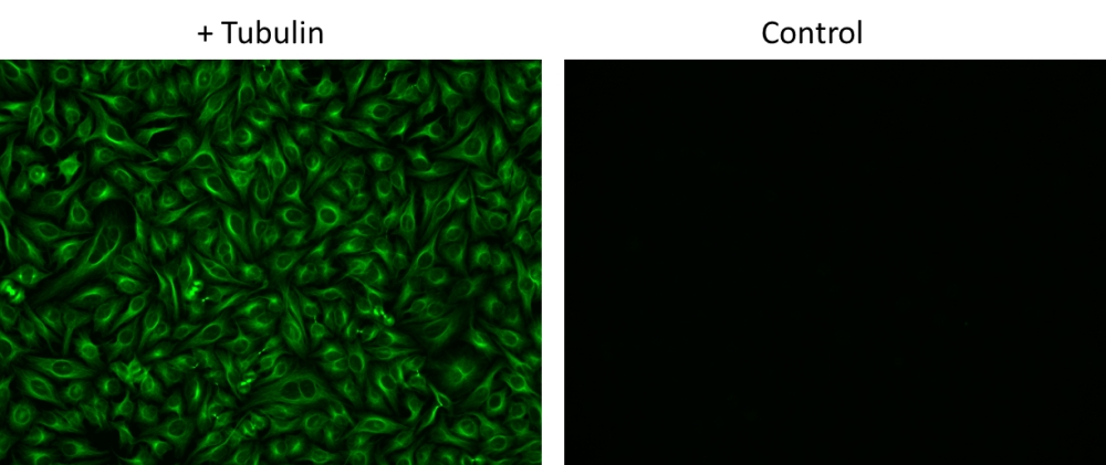
图1.分别用和不用小鼠抗微管蛋白染色的HeLa细胞,然后用ReadiLink™Rapid AF488抗体标记试剂盒(Cat#1275)制备的Alpha Fluor™488山羊抗小鼠IgG结合物的免疫荧光分析。
参考文献
A Fluorescent Probe to Unravel Functional Features of Cannabinoid Receptor CB1 in Human Blood and Tonsil Immune System Cells.
Authors: Martín-Fontecha, Mar and Angelina, Alba and Rückert, Beate and Rueda-Zubiaurre, Ainoa and Martín-Cruz, Leticia and van de Veen, Willem and Akdis, Mübeccel and Ortega-Gutiérrez, Silvia and López-Rodríguez, María Luz and Akdis, Cezmi A and Palomares, Oscar
Journal: Bioconjugate chemistry (2018): 382-389
N-terminal and central segments of the type 1 ryanodine receptor mediate its interaction with FK506-binding proteins.
Authors: Girgenrath, Tanya and Mahalingam, Mohana and Svensson, Bengt and Nitu, Florentin R and Cornea, Razvan L and Fessenden, James D
Journal: The Journal of biological chemistry (2013): 16073-84
SNAP-tag based agents for preclinical in vitro imaging in malignant diseases.
Authors: Amoury, Manal and Blume, Tobias and Brehm, Hannes and Niesen, Judith and Tenhaef, Niklas and Barth, Stefan and Gattenlöhner, Stefan and Helfrich, Wijnand and Fitting, Jenny and Nachreiner, Thomas and Pardo, Alessa
Journal: Current pharmaceutical design (2013): 5429-36
Transient focal membrane deformation induced by arginine-rich peptides leads to their direct penetration into cells.
Authors: Hirose, Hisaaki and Takeuchi, Toshihide and Osakada, Hiroko and Pujals, Sílvia and Katayama, Sayaka and Nakase, Ikuhiko and Kobayashi, Shouhei and Haraguchi, Tokuko and Futaki, Shiroh
Journal: Molecular therapy : the journal of the American Society of Gene Therapy (2012): 984-93
Chemical visualization of an attractant peptide, LURE.
Authors: Goto, Hiroaki and Okuda, Satohiro and Mizukami, Akane and Mori, Hitoshi and Sasaki, Narie and Kurihara, Daisuke and Higashiyama, Tetsuya
Journal: Plant & cell physiology (2011): 49-58
Multifunctional surface modification of gold-stabilized nanoparticles by bioorthogonal reactions.
Authors: Li, Xiuru and Guo, Jun and Asong, Jinkeng and Wolfert, Margreet A and Boons, Geert-Jan
Journal: Journal of the American Chemical Society (2011): 11147-53
Simultaneous detection of changes in protein expression and oxidative modification as a function of age and APOE genotype.
Authors: DeKroon, Robert M and Osorio, Cristina and Robinette, Jennifer B and Mocanu, Mihaela and Winnik, Witold M and Alzate, Oscar
Journal: Journal of proteome research (2011): 1632-44
Site-specific labeling of proteins for single-molecule FRET measurements using genetically encoded ketone functionalities.
Authors: Lemke, Edward A
Journal: Methods in molecular biology (Clifton, N.J.) (2011): 3-15
Transmission electron microscopy characterization of fluorescently labelled amyloid β 1-40 and α-synuclein aggregates.
Authors: Anderson, Valerie L and Webb, Watt W
Journal: BMC biotechnology (2011): 125
Distribution of nerve endings in the human dorsal radiocarpal ligament.
Authors: Tomita, Kazunari and Berger, Evelyn J and Berger, Richard A and Kraisarin, Jirachart and An, Kai-Nan
Journal: The Journal of hand surgery (2007): 466-73
